 Login / Register


- Clone
- 11F6 (See other available formats)
- Regulatory Status
- RUO
- Other Names
- Mki67, Ki67, Ki-67, MIB-1, KIA
- Isotype
- Rat IgG2b, κ
- Ave. Rating
- Submit a Review
- Product Citations
- publications

-

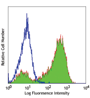
Con A+IL-2-stimulated (3 days) C57BL/6 mouse splenocytes were fixed and permeabilized with 70% ethanol, and then stained with Ki-67 (clone 11F6) Alexa Fluor® 647 (filled histogram) or rat IgG2b, κ Alexa Fluor® 647 isotype control (open histogram). -

TE-71 cells were fixed with 1% paraformaldehyde (PFA) for ten minutes, permeabilized with 0.5% Triton X-100 for ten minutes, and blocked with 5% FBS for 30 minutes. Then the cells were intracellularly stained with 2.5 µg/ml of Ki-67 (clone 11F6) Alexa Fluor® 647 (red) in 5% FBS overnight at 4°C. The cells were then stained with Phalloidin Alexa Fluor® 488 (green) for 20 minutes at room temperature. Nuclei were counterstained with DAPI (blue). The image was captured with a 20X objective. -

C57BL/6 mouse frozen intestine section was fixed with 4% paraformaldehyde (PFA) for ten minutes, permeabilized with 0.5% Triton X-100 for ten minutes, and blocked with 5% FBS plus 5% rat/mouse serum for 30 minutes at room temperature. Then the section was stained with 2.5 µg/ml of Ki-67 (clone 11F6) Alexa Fluor® 647 (red) and 5 µg/ml of E-cadherin (clone DECMA-1) Alexa Fluor® 594 (green) in 5% FBS overnight at 4°C. Nuclei were counterstained with DAPI (blue). The image was captured with a 20X objective.
| Cat # | Size | Price | Quantity Check Availability | Save | ||
|---|---|---|---|---|---|---|
| 151206 | 100 µg | 244€ | ||||
The nuclear protein Ki-67 was first identified by the monoclonal antibody Ki-67, which was generated by immunizing mice with nuclei of the L428 Hodgkin lymphoma cell line. Ki-67 protein plays an essential role in ribosomal RNA transcription and cell proliferation. Expression of Ki-67 occurs during G1, S, G2, and M phase. While in G0 phase, the Ki-67 protein is not detectable. Ki-67 is strongly expressed in proliferating cells and has been reported as a prognostic marker in various tumors.
Product DetailsProduct Details
- Verified Reactivity
- Mouse, Human
- Antibody Type
- Monoclonal
- Host Species
- Rat
- Immunogen
- E. coli expressed, N-terminal His-Thioredoxin-tagged, partial mKi-67 (1816-2163 aa) recombinant protein.
- Formulation
- Phosphate-buffered solution, pH 7.2, containing 0.09% sodium azide.
- Preparation
- The antibody was purified by affinity chromatography and conjugated with Alexa Fluor® 647 under optimal conditions.
- Concentration
- 0.5 mg/ml
- Storage & Handling
- The antibody solution should be stored undiluted between 2°C and 8°C, and protected from prolonged exposure to light. Do not freeze.
- Application
-
ICFC - Quality tested
ICC, IHC-F - Verified - Recommended Usage
-
Each lot of this antibody is quality control tested by intracellular immunofluorescent staining with flow cytometric analysis. For intracellular flow cytometric staining, the suggested use of this reagent is ≤0.125 µg per million cells in 100 µl volume. For immunocytochemistry, a concentration range of 2.5 - 5.0 μg/ml is recommended.For immunohistochemical staining on frozen tissue sections, the suggested use of this reagent is 1.25 - 5.0 µg per ml. It is recommended that the reagent be titrated for optimal performance for each application.
* Alexa Fluor® 647 has a maximum emission of 668 nm when it is excited at 633 nm / 635 nm.
Alexa Fluor® and Pacific Blue™ are trademarks of Life Technologies Corporation.
View full statement regarding label licenses - Excitation Laser
-
Red Laser (633 nm)
- Product Citations
-
- RRID
-
AB_2566801 (BioLegend Cat. No. 151206)
Antigen Details
- Structure
- 325 kD protein containing a forkhead-associated (FHA) domain and 13 tandem repeats.
- Distribution
-
Nucleus and chromosomes.
- Function
- Required for cell cycle progression and proliferation.
- Biology Area
- Cell Biology, Cell Cycle/DNA Replication
- Antigen References
-
- Starborg M, et al. 1996. J. Cell. Sci. 109:143.
- Byeon IJ, et al. 2005. Nat. Struct. Mol. Biol. 12:987.
- Yerushalmi R, et al. 2010. Lancet. Oncol. 11:174.
- Beltrami AP, et al. 2001. N. Engl. J. Med. 344:1750.
- Sachsenberg N, et al. 1998. J. Exp. Med. 187:1295.
- Nagy Z, et al. 1997. Acta. Neuropathol. 93:294.
- Gene ID
- 4288 View all products for this Gene ID 17345 View all products for this Gene ID
- UniProt
- View information about Ki-67 on UniProt.org


Other Formats
View All Ki-67 Reagents Request Custom Conjugation| Description | Clone | Applications |
|---|---|---|
| Purified anti-mouse/human Ki-67 | 11F6 | IHC-F,ICC |
| Alexa Fluor® 488 anti-mouse/human Ki-67 | 11F6 | ICFC,ICC,IHC-F |
| Alexa Fluor® 647 anti-mouse/human Ki-67 | 11F6 | ICFC,ICC,IHC-F |
| Brilliant Violet 421™ anti-mouse/human Ki-67 | 11F6 | ICFC,IHC-F |
| PE anti-mouse/human Ki-67 | 11F6 | ICFC |
| Alexa Fluor® 594 anti-mouse/human Ki-67 | 11F6 | IHC-F |
| FITC anti-mouse/human Ki-67 | 11F6 | ICFC |
| Brilliant Violet 650™ anti-mouse/human Ki-67 | 11F6 | ICFC |
| PE/Cyanine7 anti-mouse/human Ki-67 | 11F6 | ICFC |
| PE/Dazzle™ 594 anti-mouse/human Ki-67 | 11F6 | ICFC |
| PerCP/Cyanine5.5 anti-mouse/human Ki-67 | 11F6 | ICFC |
| Pacific Blue™ anti-mouse/human Ki-67 | 11F6 | ICFC |
| Brilliant Violet 711™ anti-mouse/human Ki-67 | 11F6 | ICFC |
| Brilliant Violet 510™ anti-mouse/human Ki-67 | 11F6 | ICFC |
Customers Also Purchased
Compare Data Across All Formats
This data display is provided for general comparisons between formats.
Your actual data may vary due to variations in samples, target cells, instruments and their settings, staining conditions, and other factors.
If you need assistance with selecting the best format contact our expert technical support team.
-
Purified anti-mouse/human Ki-67

TE-71 cells were fixed with 1% paraformaldehyde (PFA) for te... 
C57BL/6 mouse frozen intestine section was fixed with 4% par... -
Alexa Fluor® 488 anti-mouse/human Ki-67

TE‐71 cells were fixed with 1% paraformaldehyde (PFA) for te... 
C57BL/6 mouse frozen intestine section was fixed with 4% par... 
Con A+IL-2-stimulated (3 days) C57BL/6 mouse splenocytes wer... -
Alexa Fluor® 647 anti-mouse/human Ki-67

TE-71 cells were fixed with 1% paraformaldehyde (PFA) for te... 
C57BL/6 mouse frozen intestine section was fixed with 4% par... 
Con A+IL-2-stimulated (3 days) C57BL/6 mouse splenocytes wer... -
Brilliant Violet 421™ anti-mouse/human Ki-67

C57BL/6 frozen intestine section was fixed with 4% paraforma... 
Con A+IL-2-stimulated (3 days) C57BL/6 mouse splenocytes wer... 
Con A+IL-2-stimulated (3 days) C57BL/6 mouse splenocytes wer... -
PE anti-mouse/human Ki-67

ConA/IL-2 treated C57BL/6 mouse splenocytes were fixed and p... -
Alexa Fluor® 594 anti-mouse/human Ki-67

C57BL/6 mouse frozen intestine section was fixed with 4% par... -
FITC anti-mouse/human Ki-67

Con A+IL-2-stimulated (3 days) C57BL/6 mouse splenocytes wer... 
Con A+IL-2-stimulated (3 days) C57BL/6 mouse splenocytes we... -
Brilliant Violet 650™ anti-mouse/human Ki-67

C57BL/6J splenocytes treated with ConA and IL-2 for 2 days w... -
PE/Cyanine7 anti-mouse/human Ki-67

C57BL/6J splenocytes treated with ConA and IL-2 for 3 days w... -
PE/Dazzle™ 594 anti-mouse/human Ki-67

C57BL/6J splenocytes treated with ConA and IL-2 for 3 days w... -
PerCP/Cyanine5.5 anti-mouse/human Ki-67

C57BL/6J splenocytes treated with ConA and IL-2 for 3 days w... -
Pacific Blue™ anti-mouse/human Ki-67

C57BL/6J splenocytes treated with ConA and IL-2 for 3 days w... -
Brilliant Violet 711™ anti-mouse/human Ki-67

Con A+IL-2-stimulated (3 days) C57BL/6 mouse splenocytes wer... 
Con A+IL-2-stimulated (3 days) C57BL/6 mouse splenocytes wer... 
Con A+IL-2-stimulated (3 days) C57BL/6 mouse splenocytes wer... -
Brilliant Violet 510™ anti-mouse/human Ki-67

Con A+IL-2-stimulated (3 days) C57BL/6 mouse splenocytes wer... 
Con A+IL-2-stimulated (3 days) C57BL/6 mouse splenocytes wer... 
Con A+IL-2-stimulated (3 days) C57BL/6 mouse splenocytes wer...


Follow Us