 Login / Register


- Clone
- TS2/16 (See other available formats)
- Regulatory Status
- RUO
- Workshop
- V A-S202
- Other Names
- Integrin β1 chain, VLA-β chain, gpIIa, ITGB1
- Isotype
- Mouse IgG1, κ
- Ave. Rating
- Submit a Review
- Product Citations
- publications

-

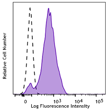
Human peripheral blood lymphocytes stained with purified TS2/16, followed by anti-mouse IgGs FITC -

BT474 breast cancer cells were stained with anti-CD29 (clone TS2/16) followed by DyLight™ 649 Goat anti-mouse Ig secondary antibody (red), plus DAPI staining for nuclei (blue). Images were taken under 20x bin4 (Filter set: EX647/10x, Dichroic 665LP, EM 700/70x) at exposure 4s. Data provided by Er Liu and John Nolan, La Jolla Institute for Bioengineering.
| Cat # | Size | Price | Quantity Check Availability | Save | ||
|---|---|---|---|---|---|---|
| 303001 | 25 µg | 48€ | ||||
| 303002 | 100 µg | 90€ | ||||
CD29 is a 130 kD single chain type I glycoprotein also known as integrin β1, VLA-β chain, or gpIIa. It is broadly expressed on a majority of hematopoietic and non-hematopoietic cells, including leukocytes (although at low level on granulocytes), platelets, fibroblasts, endothelial cells, epithelial cells, and mast cells. CD29 is a member of the integrin family. It is non-covalently associated with integrin α1-α6 chains to form VLA-1 to VLA-6 molecules, respectively. Integrins, which include CD29, bind to several cell surface (e.g. VCAM-1, MadCAM-1) and extracellular matrix molecules. CD29 acts as a fibronectin receptor and is involved in a variety of cell-cell and cell-matrix interactions.
Product DetailsProduct Details
- Verified Reactivity
- Human
- Reported Reactivity
- African Green, Baboon, Cow, Cynomolgus, Dog, Horse, Rhesus
- Antibody Type
- Monoclonal
- Host Species
- Mouse
- Formulation
- Phosphate-buffered solution, pH 7.2, containing 0.09% sodium azide.
- Preparation
- The antibody was purified by affinity chromatography.
- Concentration
- 0.5 mg/mL
- Storage & Handling
- The antibody solution should be stored undiluted between 2°C and 8°C.
- Application
-
FC - Quality tested
ICC - Verified
IHC, IP - Reported in the literature, not verified in house - Recommended Usage
-
Each lot of this antibody is quality control tested by immunofluorescent staining with flow cytometric analysis. For flow cytometric staining, the suggested use of this reagent is ≤ 0.5 µg per 106 cells in 100 µL volume or 100 µL of whole blood. It is recommended that the reagent be titrated for optimal performance for each application.
- Application Notes
-
Additional reported applications (for the relevant formats) include: immunoprecipitation3, immunohistochemical staining of acetone-fixed frozen tissue sections3,5, and activation of integrin ß14,7,8. The LEAF™ purified antibody (Endotoxin <0.1 EU/µg, Azide-Free, 0.2 µm filtered) is recommended for functional assays (Cat. No. 303010). Clone TS2/16 recognizes epitope A2.10
-
Application References
(PubMed link indicates BioLegend citation) -
- Schlossman S, et al. Eds. 1995. Leucocyte Typing V. Oxford University Press. New York.
- Gutierrez-Lopez M, et al. 2003. J. Biol. Chem. 278:208.
- Hemler ME, et al. 1984. J. Immunol. 132:3011. (IHC, IP)
- Sanchez-Aparicio P, et al. 1994. J. Cell Biol. 126:271. (Activ)
- Frank NY, et al. 2005. Cancer Res. 65:4320. (IHC)
- Murga M, et al. 2005. Blood 105:1992. (FC) PubMed
- Porter JC and Hogg N. 1997. J. Cell Biol. 138:1437. (Activ)
- Conway RE, et al. 2006. Mol. Cell. Biol. 26:5310. (Activ)
- Wesseling J, et al. 1995. J. Cell. Biol. 129:255. (Dog Reactivity)
- Rubio G, et al. 2002. Cancer Immunol. Immunother. 51:130.
- Dong A, et al. 2015. J Biol Chem. 290:8016. PubMed
- Paebst F, et al. 2014. Cytometry A. 85(8):678-87. (Horse reactivity)
- Product Citations
-
- RRID
-
AB_314317 (BioLegend Cat. No. 303001)
AB_314318 (BioLegend Cat. No. 303002)
Antigen Details
- Structure
- Integrin, type I glycoprotein, forms VLA-1 to VLA-6 heterodimers with CD49a-f (α1-α6), also associates with CD51 (αV), and α7- α9, 130 kD
- Distribution
-
Lymphocytes, monocytes, granulocytes (low), platelets, mast cells, fibroblasts, endothelial cells
- Function
- Cell-cell and cell-matrix interactions
- Ligand/Receptor
- VCAM-1, MAdCAM-1, ECM
- Cell Type
- Embryonic Stem Cells, Endothelial cells, Fibroblasts, Granulocytes, Lymphocytes, Mast cells, Mesenchymal Stem Cells, Monocytes, Platelets, Tregs
- Biology Area
- Cell Adhesion, Cell Biology, Immunology, Innate Immunity, Stem Cells
- Molecular Family
- Adhesion Molecules, CD Molecules
- Antigen References
-
1. Hemler M. 1990. Annu. Rev. Immunol. 8:365.
2. Hynes R. 1992. Cell 69:11. - Gene ID
- 3688 View all products for this Gene ID
- UniProt
- View information about CD29 on UniProt.org


Related FAQs
Other Formats
View All CD29 Reagents Request Custom Conjugation| Description | Clone | Applications |
|---|---|---|
| APC anti-human CD29 | TS2/16 | FC |
| PE anti-human CD29 | TS2/16 | FC |
| PE/Cyanine5 anti-human CD29 | TS2/16 | FC |
| Purified anti-human CD29 | TS2/16 | FC,ICC,IHC,IP |
| APC/Cyanine7 anti-human CD29 | TS2/16 | FC |
| Alexa Fluor® 488 anti-human CD29 | TS2/16 | FC |
| Alexa Fluor® 647 anti-human CD29 | TS2/16 | FC |
| Alexa Fluor® 700 anti-human CD29 | TS2/16 | FC |
| Purified anti-human CD29 (Maxpar® Ready) | TS2/16 | FC,CyTOF® |
| PerCP/Cyanine5.5 anti-human CD29 | TS2/16 | FC |
| PE/Cyanine7 anti-human CD29 | TS2/16 | FC |
| TotalSeq™-A0369 anti-human CD29 | TS2/16 | PG |
| TotalSeq™-C0369 anti-human CD29 | TS2/16 | PG |
| TotalSeq™-B0369 anti-human CD29 | TS2/16 | PG |
| PE/Dazzle™ 594 anti-human CD29 | TS2/16 | FC |
| Ultra-LEAF™ Purified anti-human CD29 | TS2/16 | FC,ICC,IHC,IP,Activ |
| TotalSeq™-D0369 anti-human CD29 | TS2/16 | PG |
Customers Also Purchased
Compare Data Across All Formats
This data display is provided for general comparisons between formats.
Your actual data may vary due to variations in samples, target cells, instruments and their settings, staining conditions, and other factors.
If you need assistance with selecting the best format contact our expert technical support team.
-
APC anti-human CD29

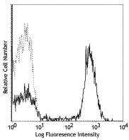
Human peripheral blood lymphocytes stained with TS2/16 APC -
PE anti-human CD29

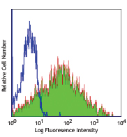
Human peripheral blood lymphocytes stained with TS2/16 PE -
PE/Cyanine5 anti-human CD29

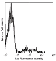
Human peripheral blood lymphocytes stained with TS2/16 PE/Cy... -
Purified anti-human CD29

Human peripheral blood lymphocytes stained with purified TS2... 
BT474 breast cancer cells were stained with anti-CD29 (clone... -
APC/Cyanine7 anti-human CD29

Human peripheral blood lymphocytes stained with TS2/16 APC/C... -
Alexa Fluor® 488 anti-human CD29

Human peripheral blood lymphocytes stained with TS2/16 Alexa... -
Alexa Fluor® 647 anti-human CD29

Human peripheral blood lymphocytes stained with TS2/16 Alexa... -
Alexa Fluor® 700 anti-human CD29

Human peripheral blood lymphocytes stained with TS2/16 Alexa... -
Purified anti-human CD29 (Maxpar® Ready)

Human PBMCs stained with 154Sm-anti-CD45 (HI30) and 156Gd-an... -
PerCP/Cyanine5.5 anti-human CD29

Human peripheral blood lymphocytes stained with CD29 (clone ... -
PE/Cyanine7 anti-human CD29

Human peripheral blood lymphocytes stained with CD29 (clone ... -
TotalSeq™-A0369 anti-human CD29
-
TotalSeq™-C0369 anti-human CD29
-
TotalSeq™-B0369 anti-human CD29
-
PE/Dazzle™ 594 anti-human CD29

Human peripheral blood lymphocytes stained with CD3 APC and ... -
Ultra-LEAF™ Purified anti-human CD29

Human peripheral blood lymphocytes stained with Ultra-LEAF™ ... -
TotalSeq™-D0369 anti-human CD29


Follow Us