Apotracker™

Calcium-independent apoptosis probes


Non-antibody probes can be particularly useful in apoptosis detection assays. Annexin V, a calcium-dependent probe, has historically been a gold standard for apoptosis detection. However, the presence of calcium in some samples may affect the viability and marker expression of cell types. For scientists looking to use calcium-independent probes that do not require a specialized buffer, we provide Apotracker™. Apotracker is a family of fluorogenic probes that bind to apoptotic cells in a calcium-independent manner, detecting externalized phosphatidylserine (PS) residues. Apotracker exhibits a linear relationship with Annexin V staining and can be useful for microscopy assays where it presents less background staining.

Apotracker™ Comparisons

We offer multiple methods for calcium-independent apoptosis detection, Apotracker Green and Apotracker Tetra. In general, directly labeled Apotracker dyes like Apotracker Green are smaller than their tetramer-based Apotracker Tetra counterparts. Because Apotracker Green only fluoresces when bound to phosphatidylserine, it is more suited for microscopy applications. Apotracker Tetra reagents create a tetramer from biotinylated monomers and report via a fluorophore-conjugated streptavidin, meaning it can potentially offer more sensitivity with the tradeoff of extra protocol steps to create the tetramer. As Apotracker Tetra is constitutively fluorescent, it is less suited for microscopy applications where it can create background signal. Explore the table below to see additional differences between the formats and determine which is better suited to your experiments.


Properties

Apotracker™ Green 

Apotracker™ Tetra 

Molecular size

Small molecules (15 aa + Fluor)

150 aa monomers + SAv-Fluor 

Reagent preparation

Direct use/directly labeled

2-step (monomer-biotin + SAv tetramer complex)

Staining in Ca2+ free buffer (FACS buffer or equivalent)

Yes

Yes

Fluorescence mechanism

Environmentally sensitive, fluorescent only when PS-bound

Constitutively fluorescent

Fluorophores

FITC directly bound to PS-sensing probe

Flexible via selection of streptavidin-fluorophore

Extracellular vesicles (EV) identification

Yes (live-cell, in vitro labeling)

Yes (in vivo labeling, ex vivo imaging) 

Live-cell imaging

Yes

No data

Retained after fixation

Yes

Yes 

Storage

-20°C after reconstitution with DMSO for up to five years

Multimer mixture can be stored at 4°C for up to two years


Apotracker™ Green

Detected in the FITC channel, Apotracker™ Green (Apo-15) is a fluorogenic probe that binds to apoptotic cells in a Ca2+ independent manner, while exhibiting a linear relationship with Annexin V staining, suggesting they are both detecting externalized PS residues. Apotracker™ Green is also useful in microscopy applications on live cells and is retained with paraformaldehyde fixation.

Identify cells in apoptosis by flow cytometry and microscopy

Apotracker™ Green (Apo-15) microscopy image


5-day old HeLa cells were stained with Calcein Red-AM (an indicator of live, healthy cells), Helix NP™ Blue, and Apotracker Green (Apo-15).

Apotracker™ Green (Apo-15) data

Day-old murine splenocytes were stained with Apotracker Green, washed and then either analyzed unfixed (black) or fixed with FluoroFix™ Buffer (red). Signal could be affected by other harsher fix and permeabilization reagents.

Apotracker™ Green labels apoptotic, dead cells without the need for specialized buffers

Apotracker™ Green (Apo-15) flow data image Unstimulated (left) and CD95-stimulated (right) Jurkat cells were stained with Apotracker Green (Apo-15) and Helix NP NIR. Cells were stained for 10-15 min in FACS buffer followed by two washes prior to analysis.


Flow data with Apotracker™ Green (Apo-15) (Left) Day-old splenocytes, gated on Helix NP NIR negative cells to exclude necrotic cells. (Right) Helix NP NIR-negative cells (red gate, left plot) stained for Apotracker Green and BV421™ Annexin V. Staining was performed with Annexin Binding Buffer according to BioLegend protocol. It has not been determined if Annexin V and Apotracker Green compete for binding sites.


To learn more, visit our Apoptosis page.


Apotracker™ Tetra

Similar to Apotracker Green, Apotracker Tetra detects phosphatidylserine accumulation on dead and apoptotic cells without the use of cation-containing specialized buffers. The reagent contains two components: biotinylated monomeric peptides and fluorophore-conjugated streptavidin. Upon mixture of the two components and formation of a tetramer complex, the peptides undergo conformational changes, resulting in higher affinity binding of the peptide components to phosphatidylserine residues. This enables specific detection of dead and apoptotic cells, as well as extracellular vesicles.


Apotracker Tetra is currently available as a product bundle that includes the biotinylated monomer peptide and streptavidin-Alexa Fluor® 647, which is pre-diluted and titrated for ease of use. The biotinylated monomer components can then be paired with any available streptavidin-fluorophore option, providing additional flexibility in multicolor panel construction. For guidance on titration of your streptavidin conjugate, you can contact Technical Services


Apotracker™ Tetra staining correlates with other apoptotic cell indicators

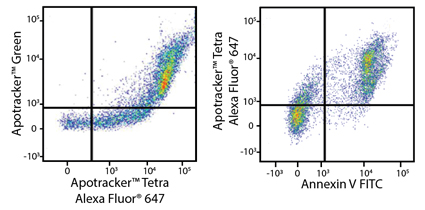
Heat-induced C57BL/6 splenocytes, containing 40% dead cells (left) and 50% dead cells (right) stained with Apotracker Green and Apotracker Tetra Alexa Fluor® 647. Signal of Apotracker Green and Apotracker Tetra Alexa Fluor® 647 showed a positive correlation (left).


Apotracker™ Tetra stains both mouse splenocytes and human PBMCs


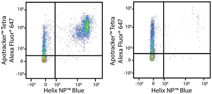
Heat-induced 50% dead C57BL/6 splenocytes (left) or untreated splenocytes (right) stained with Helix NP Blue and Apotracker Tetra Alexa Fluor® 647.


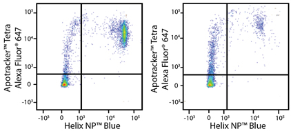
Heat-induced 50% dead PBMC (left) or untreated PBMC (right) stained with Helix NP Blue and Apotracker Tetra Alexa Fluor® 647.

ProductsHere

Login / Register
Remember me
Forgot your password? Reset password?
Create an Account