 Login / Register


- Clone
- 14G10A21 (See other available formats)
- Regulatory Status
- RUO
- Other Names
- Nuclear factor NF-kappa-B p65 subunit (NF-κB p65), Rel-A, transcription factor p65, Nuclear factor of kappa light polypeptide gene enhancer in B-cells 3 (NFKB3)
- Isotype
- Mouse IgG2b, κ
- Ave. Rating
- Submit a Review
- Product Citations
- publications

-

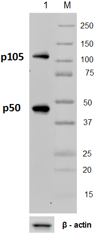
Total lysates (15µg protein) from 293T and 293T/NF-κB p65 knockdown(KD) cells were resolved by electrophoresis (4-20% Tris-Glycine gel), transferred to nitrocellulose, and probed with 1:500 diluted (1µg/mL) purified anti-NF-κB p65 antibody, clone 14G10A21 (upper) or 1:3000 diluted anti-GAPDH (poly6314) antibody (lower). Proteins were visualized by chemiluminescence detection using a 1:3000 diluted goat anti-mouse-IgG secondary antibody conjugated to HRP for the anti-NF-κB p65 antibody, and a donkey anti-rabbit IgG Antibody conjugated to HRP for GAPDH. Lane M: Molecular weight ladder, M* indicates longer exposure. -

Hela cell extracts were resolved by electrophoresis, transferred to nitrocellulose, and probed with purified monoclonal anti-NF-κB p65 (clone 14G10A21) antibody. Proteins were visualized using an anti-mouse-IgG secondary conjugated to HRP and chemiluminescence detection. -

Immunoprecipitation of NF-κB p65 from Hela cell extracts. Lane 1 is 5% input. Immunoprecipitation was performed using protein G resins only (lane 2), mouse IgG isotype control (lane 3), and anti-NF-κB p65 antibody (clone 14G10A21, lane 4). Western blot was performed using anti-NF-κB p65 antibody (clone 14G10A21).
| Cat # | Size | Price | Quantity Check Availability | Save | ||
|---|---|---|---|---|---|---|
| 653001 | 25 µg | £85 | ||||
| 653002 | 100 µg | £182 | ||||
NF-κB p65 is a member of REL-like domain containing protein family, which forms a NF-κB complex with the other family members: NF-κB1 (p105/p50) or NF-κB2 (p100/p52). The NF-κB complex is inactivated and held in the cytoplasm by the NF-κB inhibitor IκB. In response to activation stimuli, IκB kinases (IKKs) phosphorylates IκB, resulting in degradation of IκB and liberation of NF-κB complex. The activated NF-κB complex translocates to the nucleus and binds to κB sites in the DNA of their target genes.
Product DetailsProduct Details
- Verified Reactivity
- Human
- Antibody Type
- Monoclonal
- Host Species
- Mouse
- Immunogen
- Partial human NF-κB p65 recombinant protein (451-551 aa)
- Formulation
-
This antibody is provided in phosphate-buffered solution, pH 7.2, containing 0.09% sodium azide.
Previous lots of this product may have been formulated with 0.1% or 0.05% NaN3 instead of 0.09% NaN3. For further information please contact BioLegend Technical Support or Customer Service. - Concentration
- 0.5 mg/ml
- Storage & Handling
- Upon receipt, store undiluted between 2°C and 8°C.
- Application
-
WB - Quality tested
IP, KO/KD-WB - Verified - Recommended Usage
-
Each lot of this antibody is quality control tested by Western blotting. For Western blotting, the suggested use of this reagent is 0.1-1.0 µg per ml. For immunoprecipitation, the suggested use of this reagent is 2-10 μg per ml. It is recommended that the reagent be titrated for optimal performance for each application.
- Application Notes
-
This clone is not recommended for ChIP (Chromatin Immunoprecipitation) assays (as determined by in-house testing).
- Product Citations
-
- RRID
-
AB_2561612 (BioLegend Cat. No. 653001)
AB_2561613 (BioLegend Cat. No. 653002)
Antigen Details
- Structure
- 65 kD protein containing a Rel homology domain (RHD), an activation domain, and a 9aaTAD domain.
- Distribution
-
The inactivated NF-κB complex containing p65 subunit is bound to IκB and is localized to cytoplasm. Upon activation, I-κB is phosphorylated and degraded. The activated NF-κB complex is in turn translocated to the nucleus as a transcription factor.
- Function
- NF-κB is a homodimeric or heterodimeric complex formed by the Rel-like domain-containing proteins. The most abundant form is p65 (RelA) - p50 (NF-κB1) heterodimer complex. The NF-κB complex is a ubiquitously expressed transcription factor which is involved in various biological functions, such as cell growth, tumorigenesis, differentiation, apoptosis, inflammation, and immune responses.
- Interaction
- Interacts with NF-κB1 (p105/p50) or NF-κB2 (p100/p52) to form heterodimeric NF-κB complex. Interacts with HDAC1, HDAC3, and CBP. Interaction with MEN1 inhibits transactivation activity of NF-κB complex.
- Cell Type
- B cells
- Biology Area
- Apoptosis/Tumor Suppressors/Cell Death, Cell Biology, Immunology, Neuroscience, Neuroscience Cell Markers, Signal Transduction, Transcription Factors
- Molecular Family
- Nuclear Markers
- Antigen References
-
1. Li Z, et al. 1997. Mol. Cell. Biol. 17:6184.
2. Saccani S, et al. 2004. J. Exp. Med. 200:107.
3. Nolan GP, et al. 1991. Cell 64:961.
4. Chen LF, et al. 2001. Science 293:1653.
5. Hansen SK, et al. 1994. Mol. Cell. Biol. 14:2593.
6. Chapman NR, et al. 2002. Biochem J. 366:459. - Gene ID
- 4790 View all products for this Gene ID
- UniProt
- View information about NF-kappaB p65 on UniProt.org

Related FAQs
Other Formats
View All NF-κB p65 Reagents Request Custom Conjugation| Description | Clone | Applications |
|---|---|---|
| Purified anti-NF-κB p65 | 14G10A21 | WB,IP,KO/KD-WB |
| PE anti-NF-κB p65 | 14G10A21 | ICFC |
| APC anti-NF-κB p65 | 14G10A21 | ICFC |
Customers Also Purchased
Compare Data Across All Formats
This data display is provided for general comparisons between formats.
Your actual data may vary due to variations in samples, target cells, instruments and their settings, staining conditions, and other factors.
If you need assistance with selecting the best format contact our expert technical support team.
-
Purified anti-NF-κB p65

Hela cell extracts were resolved by electrophoresis, transfe... 
Immunoprecipitation of NF-κB p65 from Hela cell extracts. La... 
Total lysates (15µg protein) from 293T and 293T/NF-κB p65 kn... -
PE anti-NF-κB p65

Human lung adenocarcinoma epithelial cell line A549 was fixe... -
APC anti-NF-κB p65

Human lung adenocarcinoma epithelial cell line A549 was fixe...


Follow Us