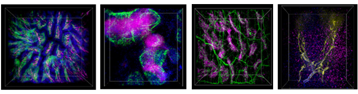
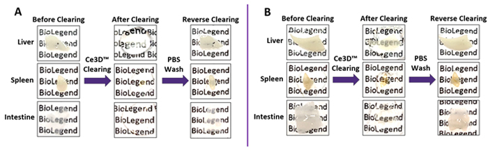
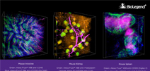
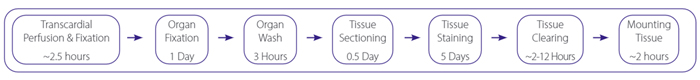
Biological tissues are generally composed of proteins, lipids, and water, each of which has a different refractive index (RI). Differences in refractive indices, or RI mismatch, cause the scattering of light in the tissue and result in tissue opacity. Tissue clearing reagents aim to reduce light scattering by normalizing the RI throughout the tissue, thus making the tissue transparent. Optical clearing is a valuable application that allows us gain a better understanding of spatial composition, phenotypic and subtype identity, and cellular networks. Our Ce3D™ Tissue Clearing Solution pairs perfectly with our expansive portfolio of antibodies, generating mesmerizing 3D images with unparalleled depth.


The Ce3D™ tissue clearing system was developed by the Laboratory of Immune System Biology (LISB) in the National Institute of Allergy and Infectious Diseases (NIAID, NIH). The original methods can be reviewed below:


Tutorial: See 3D (Ce3D™): Enhanced Tissue Clearing for Multicolor Microscopy


Watch our video explaining the capabilities of Ce3D™.

3D IHC Data


3D Imaging


The images above are representative 3D IHC images of 500 μm-thick mouse samples. The sections were optically cleared after immunostaining, mounted in sample chambers, and imaged with a 10X or 20X objective.

From left to right: 

Watch dozens of our 3D imaging videos.


Optical Clearing Using Ce3D™ Tissue Clearing Solution


Representative 500 µm-thick mouse tissue sections before and after tissue clearing. Mice were perfused and organs were fixed with 1% (A) or 4% (B) PFA. Clearing was reversed by immersing tissues in PBS for one hour. 1% PFA is recommended for best clearing effect. 4% PFA is recommended for optimal epitope preservation and immunostaining.


Optical Clearing Using Ce3D™ Tissue Clearing Solution

What is Ce3D™?


Ce3D™ (clearing-enhanced 3D) Tissue Clearing Reagent is a hydrophilic and RI-matching solution, making it easy-to-use for the rapid and robust clearing of diverse tissues. Ce3D™ has an RI of ~1.5 and is compatible with multicolor fluorescence microscopy. The Ce3D™ tissue clearing technique works by simple immersion of tissue into the clearing reagent, and does not require special equipment or containers. Ce3D™ offers many advantages:


Key Features


  • Simple, one-step passive clearing process
  • Preservation of fluorescent signal, tissue architecture, and cell morphology
  • Reversible to allow 2D IHC and other downstream applications
  • Available as pre-assembled kits or as individual reagents, saving time on buffer preparation
  • Validated with over 60 BioLegend antibody clones and conjugated formats

Ce3D™ Tissue Clearing Kits


Test Size Cat. No. Ce3D™ Tissue Clearing Solution Ce3D™ Permeabilization/Blocking Buffer Ce3D™ Antibody Diluent Buffer Ce3D™ Wash Buffer
10 Tests 427701 5 mL 15 mL 15 mL 50 mL
50 Tests 427702 25 mL 50 mL 50 mL 250 mL


Ce3D™ Tissue Clearing Individual Reagents


Reagent Size (µL) Cat. No.
Ce3D™ Tissue Clearing Solution 5 mL | 25 mL 427703 | 427704
Ce3D™ Permeabilization/Blocking Buffer 25 mL | 50 mL 427705 | 427706
Ce3D™ Antibody Diluent Buffer 15 mL | 50 mL 427707 | 427708
Ce3D™ Wash Buffer 50 mL | 250 mL 427709 | 427710

Ce3D™ Tissue Clearing Kit Staining Protocol for Microscopy


View our step-by-step protocol video
for Ce3D™ reagents.

Our protocol and troubleshooting guide provides an in-depth optimized protocol, complete with notes and commentary for each step. The guide also outlines the entire procedure, leading you through perfusion, fixation, sectioning, staining, tissue clearing, and mounting steps. The process itself can typically be completed within 7 days.


In addition, the guide contains suggestions for resolving potential issues you may encounter as you optimize your experiment. Read through it for tips on fixation, uneven staining, and high background. For additional information or help, contact our Technical Services team.

Recommended Clearing Time for Different Tissues


Time Tissue
2 hours Intestine, lymph node
4 hours Spleen, thymus, lung
> 8 hours Kidney, liver, testis, brain

Ce3D™ Tissue Clearing Workflow


Below is a list of the antibodies we have validated in-house with our Ce3D™ Tissue Clearing Solution. We will continue to test and expand this list as new conjugates become available.


Validated Anti-Mouse Antibodies for Ce3D™ Tissue Clearing


Specificity Clone Alexa Fluor® 488 Alexa Fluor® 594 Alexa Fluor® 647
CD3 17A2 X X X
CD3ε 145-2C11     X
CD3ε KT3.1.1     X
CD3ε 500A2 X X X
CD4 RM4-5 X   X
CD8a  53-6.7 X X X
CD8a C8/144B     X
CD11b M1/70 X X X
CD11c N418   X X
CD18 M18/2   X X
CD21/CD35 (CR2/CR1) 7E9   X X
CD22 OX-97     X
CD31 MEC13.3   X X
CD37 Duno85   X  
CD45 30-F11 X X X
CD45R/B220 RA3-6B2 X X X
CD68 FA-11 X X X
CD106 429 (MVCAM.A)     X
CD117 (c-Kit) 2B8   X  
CD146 ME-9F1   X X
CD147 OX-114 X    
CD169 (Siglec-1) 3D6.112 X X X
CD172a (SIRPα) P84   X X
CD200 (OX2) OX-90   X X
CD324 (E-Cadherin) DECMA-1   X X
CD326 (Ep-CAM) G8.8 X X X
CLEC4F 3E3F9     X
F4/80 BM8 X   X
I-A/I-E M5/114.15.2 X   X
IgD 11-26c.2a X X X
Podoplanin PMab-1 X X X
Tubulin β 3 (TUBB3) TUJ1 X X X
ProductsHere

Login / Register
Remember me
Forgot your password? Reset password?
Create an Account