 Login/Register


Neurons are highly specialized cells with unique compartments that are distinguishable using specific markers. These compartments can be generally classified into soma (cell body), axon, dendrite, and synapse. Certain markers allow for discrimination of neurons from other cell types in the nervous system, namely microglia, astrocytes, and oligodendrocytes (ODs). The use of antibodies for these markers in conjunction with microscopy serves as a powerful method for collecting data relevant for, but not limited to: 1) cell type identification, 2) cellular co-localization, 3) phenotypic and morphological analysis, and 4) protein expression levels. To this end, BioLegend offers a number of antibodies against cell type specific and structural markers that have been validated for use in IHC and/or ICC. Our Neuron Marker Antibody Sampler Kit includes antibodies for markers expressed in different compartments of a neuron.
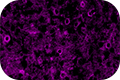 Clone NSE-P1 detects Enolase 2, also known as NSE, which is a soluble protein used for identification of neurons and cells of neuronal origin. This antibody can be used to visualize the soma and neuronal processes.
 Clone 1B7 was raised against human Neuronal Nuclei (NeuN) protein, also known as Fox3. This antibody reveals strong nuclear staining of a wide range of neuronal cell types. There are some neuronal cells that are not detected by NeuN, such as Purkinje neurons, Golgi cells, and retinal photoreceptor cells.
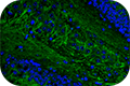 Clone SMI 52 reacts with the structural protein microtubule-associated protein 2 (MAP2), and recognizes neuronal cell bodies and dendrites in tissue sections and cell cultures.
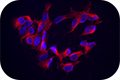 Clone TUJ1 is highly reactive to class III β-tubulin, another cytoskeletal protein expressed in neurons. Clone TUJ1 does not react with β-tubulin found in glial cells. Immunostaining with TUJ1 allows visualization of cell bodies, dendrites, and axons.
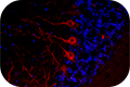 Clone SMI 32 detects neurofilaments (NF), which are a major component of the neuronal cytoskeleton. There are three major NF subunits distinguished by mass – light (NF-L), medium (NF-M), and heavy (NF-H). Immunostaining with SMI 32 detects NF-H and is useful for discriminating neurons (NF-positive) from glia (NF-negative).


Follow Us