 Login/Register


- Clone
- BSB-62 (See other available formats)
- Regulatory Status
- RUO
- Other Names
- Transcription factor SOX-10
- Isotype
- Mouse IgG2b, κ
- Ave. Rating
- Submit a Review
- Product Citations
- publications

-

IHC staining of purified anti-SOX-10 antibody (clone BSB-62) on formalin-fixed paraffin-embedded rat brain tissue. Following antigen retrieval using Sodium Citrate H.I.E.R., the tissue was incubated with 5 µg/mL of the primary antibody overnight at 4°C, followed by incubation with Alexa Fluor® 594 goat anti-Mouse IgG (Cat. No. 405326) for one hour at room temperature. Nuclei were counterstained with DAPI. The image was captured with a 40X objective. Scale bar: 50 µm. -

IHC staining of purified anti-SOX-10 antibody (clone BSB-62) on formalin-fixed paraffin-embedded mouse brain tissue. Following antigen retrieval using Sodium Citrate H.I.E.R., the tissue was incubated with 5 µg/mL of the primary antibody overnight at 4°C, followed by incubation with Alexa Fluor® 594 goat anti-Mouse IgG (Cat. No. 405326) for one hour at room temperature. Nuclei were counterstained with DAPI. The image was captured with a 40X objective. Scale bar: 50 µm. -

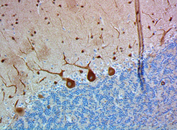
IHC staining of purified anti-SOX-10 antibody (clone BSB-62) on formalin-fixed paraffin-embedded rat and mouse brain tissue. Following antigen retrieval using Sodium Citrate H.I.E.R., the tissue was incubated with 5 µg/mL of the primary antibody overnight at 4°C. BioLegend's Ultra-Streptavidin (USA) HRP kit (Multi-Species, DAB, Cat. No. 929901) was used for detection followed by hematoxylin counterstaining, according to the protocol provided. The images were captured with a 40X objective. Scale bar: 50 µm -

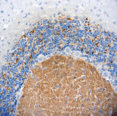
IHC staining of anti-SOX-10 antibody (clone BSB-62) on formalin-fixed paraffin-embedded human glial tumor tissue. Following antigen retrieval using Sodium Citrate H.I.E.R., the tissue was incubated with 10 µg/mL of the primary antibody for 30 minutes at room temperature. BioLegend's Ultra-Streptavidin (USA) HRP Detection Kit (Multi-Species, DAB, Cat. No. 929901) was used for detection followed by hematoxylin counterstaining, according to the protocol provided. The images were captured with a 40X objective.
| Cat # | Size | Price | Quantity Check Availability | Save | ||
|---|---|---|---|---|---|---|
| 847201 | 25 µg | $124 | ||||
| 847202 | 100 µg | $311 | ||||
SOX-10 is a transcriptional activator that is involved in the regulation of embryonic development and in the determination of cell fate.
Product DetailsProduct Details
- Verified Reactivity
- Human, Mouse, Rat
- Antibody Type
- Monoclonal
- Host Species
- Mouse
- Immunogen
- Synthetic peptide corresponding to the N-terminus of human SOX-10.
- Formulation
- Phosphate-buffered solution, pH 7.2, containing 0.09% sodium azide.
- Preparation
- The antibody was purified by affinity chromatography.
- Concentration
- 0.5 mg/ml
- Storage & Handling
- The antibody solution should be stored undiluted between 2°C and 8°C.
- Application
-
IHC-P - Quality tested
SB - Community verified - Recommended Usage
-
Each lot of this antibody is quality control tested by formalin-fixed paraffin-embedded immunohistochemical staining. For immunohistochemistry, a concentration range of 5.0 - 10 μg/mL is suggested. It is recommended that the reagent be titrated for optimal performance for each application.
- Application Notes
-
This antibody may show weaker reactivity to human tissues.
- Additional Product Notes
-
This product has been verified for IHC-F (Immunohistochemistry - frozen tissue sections) on the NanoString GeoMx® Digital Spatial Profiler. The GeoMx® enables researchers to perform spatial analysis of protein and RNA targets in FFPE and fresh frozen human and mouse samples. For more information about our spatial biology products and the GeoMx® platform, please visit our spatial biology page.
- Product Citations
-
- RRID
-
AB_2734617 (BioLegend Cat. No. 847201)
AB_2629666 (BioLegend Cat. No. 847202)
Antigen Details
- Structure
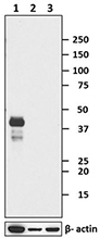
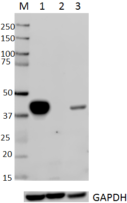
- SOX-10 is a 466 amino acid protein with a molecular mass of 50 kD.
- Distribution
-
Tissue distribution: primarily expressed in the nuclei of glia in fetal and adult brain, and in the heart, salivary gland and skin.
Cellular distribution: nucleus. - Function
- SOX-10 likely functions synergistically with the POU domain protein TST-1/OCT6/SCIP to confer function of other transcription factors in developing and mature glia. The protein acts as a nucleocytoplasmic shuttle protein and is important for neural crest and peripheral nervous system development.
- Interaction
- DNA, PAX3, NMI.
- Cell Type
- Microglia
- Biology Area
- Cell Biology, Neuroscience, Neuroscience Cell Markers
- Molecular Family
- Endosomal Markers
- Antigen References
-
1. Bondurand N, Sham MH. 2013. Dev. Biol. 382(1):330. PubMed
2. Mollaaghababa R, Pavan WJ. 2003. Oncogene. 22(20):3024. PubMed - Gene ID
- 6663 View all products for this Gene ID
- UniProt
- View information about SOX-10 on UniProt.org

Related FAQs
- If an antibody clone has been previously successfully used in IBEX in one fluorescent format, will other antibody formats work as well?
-
It’s likely that other fluorophore conjugates to the same antibody clone will also be compatible with IBEX using the same sample fixation procedure. Ultimately a directly conjugated antibody’s utility in fluorescent imaging and IBEX may be specific to the sample and microscope being used in the experiment. Some antibody clone conjugates may perform better than others due to performance differences in non-specific binding, fluorophore brightness, and other biochemical properties unique to that conjugate.
- Will antibodies my lab is already using for fluorescent or chromogenic IHC work in IBEX?
-
Fundamentally, IBEX as a technique that works much in the same way as single antibody panels or single marker IF/IHC. If you’re already successfully using an antibody clone on a sample of interest, it is likely that clone will have utility in IBEX. It is expected some optimization and testing of different antibody fluorophore conjugates will be required to find a suitable format; however, legacy microscopy techniques like chromogenic IHC on fixed or frozen tissue is an excellent place to start looking for useful antibodies.
- Are other fluorophores compatible with IBEX?
-
Over 18 fluorescent formats have been screened for use in IBEX, however, it is likely that other fluorophores are able to be rapidly bleached in IBEX. If a fluorophore format is already suitable for your imaging platform it can be tested for compatibility in IBEX.
- The same antibody works in one tissue type but not another. What is happening?
-
Differences in tissue properties may impact both the ability of an antibody to bind its target specifically and impact the ability of a specific fluorophore conjugate to overcome the background fluorescent signal in a given tissue. Secondary stains, as well as testing multiple fluorescent conjugates of the same clone, may help to troubleshoot challenging targets or tissues. Using a reference control tissue may also give confidence in the specificity of your staining.
- How can I be sure the staining I’m seeing in my tissue is real?
-
In general, best practices for validating an antibody in traditional chromogenic or fluorescent IHC are applicable to IBEX. Please reference the Nature Methods review on antibody based multiplexed imaging for resources on validating antibodies for IBEX.
Other Formats
View All SOX-10 Reagents Request Custom Conjugation| Description | Clone | Applications |
|---|---|---|
| Purified anti-SOX-10 | BSB-62 | IHC-P,SB |
Customers Also Purchased
Compare Data Across All Formats
This data display is provided for general comparisons between formats.
Your actual data may vary due to variations in samples, target cells, instruments and their settings, staining conditions, and other factors.
If you need assistance with selecting the best format contact our expert technical support team.


Follow Us