Spark Blue 550

Spark Blue™ 550


Spark Blue™ 550 provides a unique option for adding an additional fluorophore to a high-parameter (>22 color) multicolor panel on spectral unmixing cytometers like the Cytek™ Aurora or SONY ID7000™ Spectral Cell Analyzer. It is excited off of the 488 nm laser and emits at 540 nm, filling the spectral gap between commonly used fluors like FITC and PE (R-Phycoerythrin). Emission off of the 405 nm violet laser further adds to the accuracy with which it can be unmixed from neighboring fluorophores and does not significantly impact the detection of any violet laser fluorophores. Because Spark Blue™ 550 is a relatively dim fluorophore, it is best used to detect a high abundance antigen.


Excitation and Emission Spectra of Spark Blue™ 550


Spark Blue 550 spectra signature

Spark Blue 550 aurora spectra

Spark Blue 550 spectra


Emission spectra (top) and normalized emission spectra (middle) of Spark Blue™ 550 run on a 5-laser Cytek™ Aurora Spectral Cytometer. To compare Spark Blue™ 550 with other fluorophores on a spectral cytometer, use our Aurora Spectral Analyzer tool.


Normalized excitation and emission spectra (bottom) of Spark Blue™ 550 obtained from a spectrophotometer. To compare Spark Blue™ 550 with other fluorophores, use our Fluorescence Spectra Analyzer tool.

Minimal Spillover Considerations


Spark Blue™ 550 does not exhibit high spillover with other fluorophores, making it easy to place into a large panel. It is maximally excited by the 488 nm laser with minimal emission off of the 561 nm laser. While emission off of the 405 nm violet laser allows it to be easily unmixed from neighboring fluors, it does not significantly impact spreading into the violet laser fluors. Spectrally, it falls between the emissions of FITC and PE, but can easily be used in the same panel as these fluors with minimal spillover considerations.


Spark Blue 550

Spreading impact of Spark Blue™ 550 into detection channels of a 5-laser Cytek™ Aurora Spectral Cytometer.


Human whole blood was stained with anti-CD19 Spark Blue™ 550, anti-CD4 BV570™, anti-CD38 PE, and anti-CD40 FITC antibodies. Samples were unmixed on a Cytek™ Aurora Cytometer using compensation beads and cells. All plots are gated on lymphocytes.

A Brighter Alternative to Alexa Fluor® 532


Spark Blue™ 550 is spectrally similar to Alexa Fluor® 532 with a peak emission in the B3 channel on the Cytek™ Aurora and offers a brighter signal when compared to Alexa Fluor® 532 in a side-by-side experiment. Due to its unique emission peak off of the violet laser, Spark Blue™ 550 is typically easier to unmix from neighboring fluors when compared to Alexa Fluor® 532.


Spark Blue 550 compared to Alexa Fluor 532

Human peripheral blood lymphocytes were stained with anti-CD8 Pacific Blue™, and either anti-CD4 (clone SK3) Spark Blue™ 550 (left) or anti-CD4 (clone SK3) Alexa Fluor® 532 (right).

Stability and Validation Testing


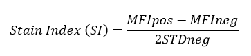
All BioLegend fluorophores undergo rigorous testing procedures to determine how light, heat, and fixation may affect the performance and ensure they will perform reliably. To compare the signal across different conditions and timepoints, we used the Stain Index (formula below) to measure the relative brightness of the antibody. 

Spark 550

Titration of Spark Blue™ 550

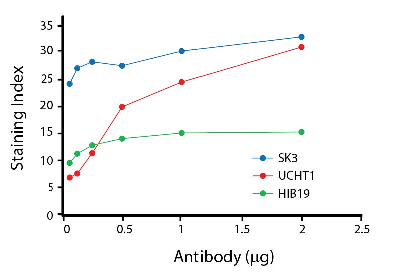
Titration curves generated by staining human lysed whole blood with Spark Blue™ 550 conjugated anti-CD4 (SK3), anti-CD3 (UCHT1), and anti-CD19 (HIB19) antibodies.


Photostability Testing


The photostability of Spark Blue™ 550 was tested in two ways that mimic how an antibody may be exposed to light over the course of an experiment.


  1. Antibodies were stored in the dark or exposed to fluorescent lighting. Then, the antibodies were used to stain freshly harvested cell samples and analyzed immediately.
  2. Cells were stained with antibody that had been kept under recommended storage conditions. Prior to analysis, the stained cells were stored in the dark or exposed to fluorescent lighting.


Spark Blue™ 550 antibodies are stable when left under fluorescent lighting overnight. When stained cells are stored overnight in either condition, there is an accelerated loss of signal due to the additive oxidative stress from the cells on the reagent.


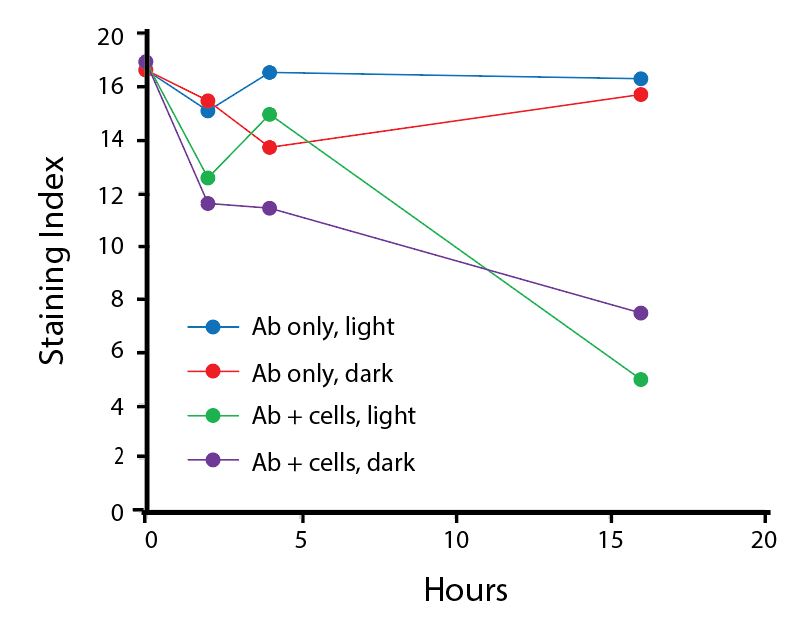
Anti-human CD4 (clone SK3) Spark Blue™ 550 was stored in a clear vial (Ab only) and left exposed to light or protected in the dark, as indicated. Antibodies were stored for the indicated timepoints and then used to stain human lysed whole blood. Samples labeled Ab+ cells contain human lysed whole blood that was stained with anti-human CD4 Spark Blue™ 550. Stained cells were then left in the light or protected, as indicated.


Heat Stability


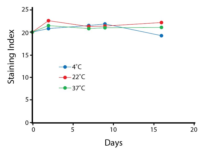
Anti-human CD4 (clone SK3) Spark Blue™ 550 was aliquoted and incubated at the indicated temperatures over the course of 17 days. The antibodies were then used to stain human lysed whole blood from a single donor.


Fixative Stability


Spark Blue™ 550 is compatible with all BioLegend fixation buffers. For each buffer set, fresh fixed samples were tested immediately following staining or stored overnight in Cyto-Last™ Buffer before being read on a cytometer.


A guide to the fixatives used in this experiment:


Spark Blue 550

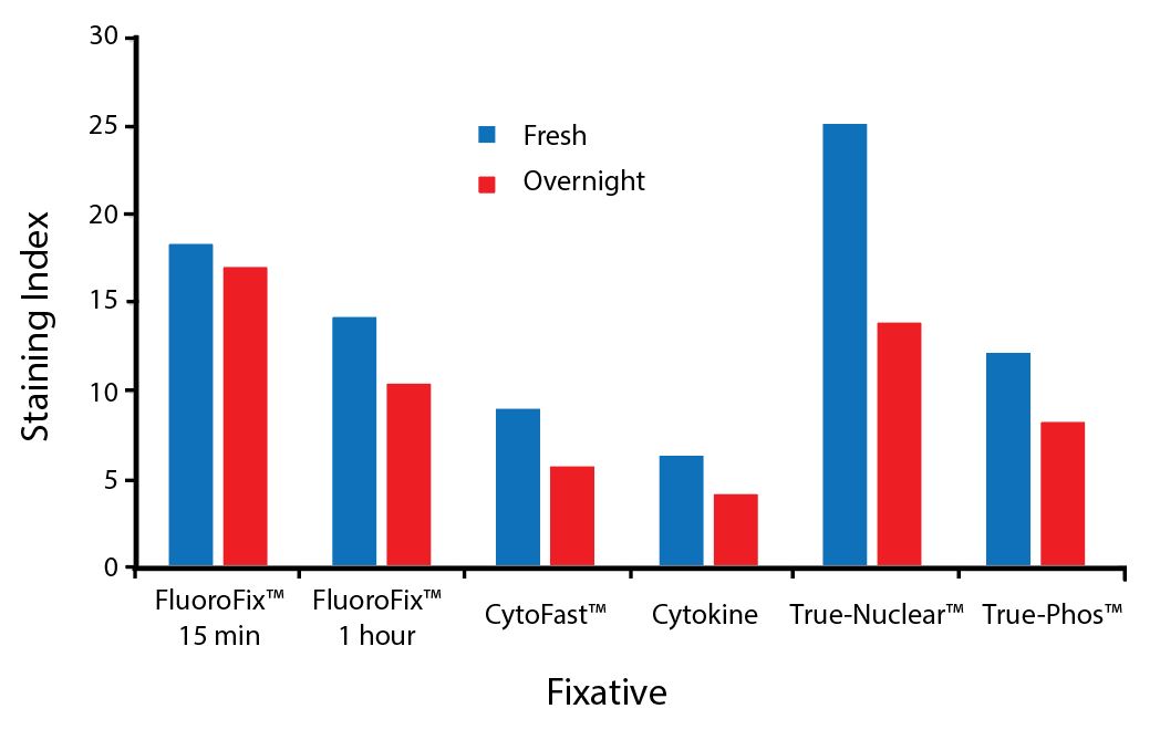
Human PBMCs were stained with anti-human CD4 (clone SK3) conjugated to Spark Blue™ 550 and fixed using the respective protocols for each buffer set. Fresh samples were fixed and read on a Cytek® Aurora immediately. Overnight samples were fixed and stored overnight in Cyto-Last™ Buffer before reading.

ProductsHere

Login/Register
Remember me
Forgot your password? Reset Password
Request an Account