 Login / Register


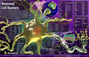
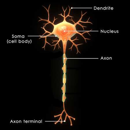
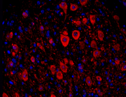
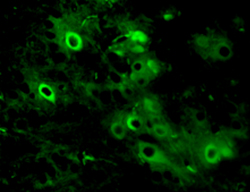
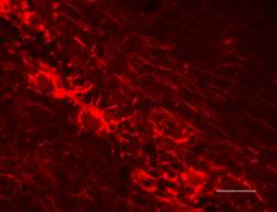
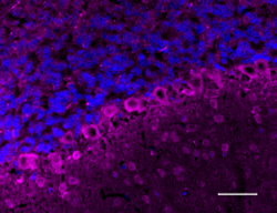
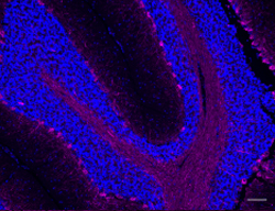
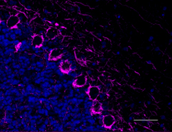
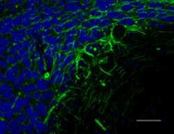
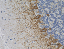
Neurons have highly compartmentalized structures that allow their distinction from other cell types in the nervous system. These unique compartments are distinguishable using specific markers, and are generally classified into soma (cell body), axon, dendrite, and synapse. The use of antibodies for these markers in conjunction with microscopy serves as a powerful method for:
|
|
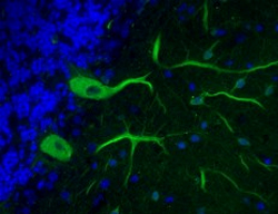
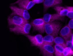
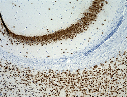
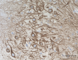
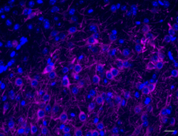
Immunolabeling of cells (immunocytochemistry or ICC) or tissues (immunohistochemistry or IHC) with antibodies to study neurons is a highly utilized application in Neuroscience mainly due to the availability of a wide range of markers and the relatively low cost for performing and imaging the immunolabeled material. Generally, target detection can be accomplished using chromogenic or fluorescent staining.


Follow Us