 Login / Register


- Clone
- MC-480 (See other available formats)
- Regulatory Status
- RUO
- Other Names
- Stage-Specific Embryonic Antigen 1, X-hapten, Lewis X, 3-FAL, CD15, SSEA1, SSEA-1
- Isotype
- Mouse IgM, κ
- Ave. Rating
- Submit a Review
- Product Citations
- publications

-

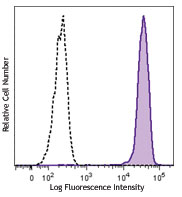
Murine embryonic carcinoma cell line F9 was stained with CD15 (SSEA-1) (clone MC-480) APC (filled histogram) or mouse IgM APC Isotype control (open histogram).
| Cat # | Size | Price | Quantity Check Availability | Save | ||
|---|---|---|---|---|---|---|
| 125617 | 25 tests | 149 CHF | ||||
| 125618 | 100 tests | 314 CHF | ||||
The MC-480 antibody reacts with mouse and human Stage-Specific Embryonic Antigen-1 (SSEA-1). It is a Lewis blood group related carbohydrate antigen, also known as X-hapten, Lewis X, 3-FAL, 3-fucosyl-N-acetyllactosamine, or CD15. The expression pattern of SSEA-1 antigen is different in humans than in mice. In mice, SSEA-1 is expressed on embryonic stem cells (ES), embryonal carcinoma cells (EC), 8-cell to blastocyst embryos, and a subset of embryonic inner cell mass. The expression on murine ES cells is decreased upon differentiation. In humans, however, SSEA-1 is not found on undifferentiated ES cells, but its expression is upregulated along with differentiation. CD15 is highly expressed on adult human granulocytes. It has been reported that SSEA-1 plays a role in cell adhesion and regulation of cell differentiation.
Product DetailsProduct Details
- Verified Reactivity
- Human, Mouse
- Antibody Type
- Monoclonal
- Host Species
- Mouse
- Immunogen
- Mouse F9 Teratocarcinoma Stem Cells (X-irradiated)
- Formulation
- Phosphate-buffered solution, pH 7.2, containing 0.09% sodium azide and BSA (origin USA)
- Preparation
- The antibody was purified by affinity chromatography and conjugated with APC under optimal conditions.
- Concentration
- Lot-specific (to obtain lot-specific concentration and expiration, please enter the lot number in our Certificate of Analysis online tool.)
- Storage & Handling
- The antibody solution should be stored undiluted between 2°C and 8°C, and protected from prolonged exposure to light. Do not freeze.
- Application
-
FC - Quality tested
- Recommended Usage
-
Each lot of this antibody is quality control tested by immunofluorescent staining with flow cytometric analysis. For flow cytometric staining, the suggested use of this reagent is 5 µl per million cells in 100 µl staining volume or 5 µl per 100 µl of whole blood.
- Excitation Laser
-
Red Laser (633 nm)
- Application Notes
-
Additional reported applications (for the relevant formats) include: immunoprecipitation1, Western blotting1, and immunohistochemistry1 of acetone-fixed frozen tissue sections and formalin-fixed paraffin-embedded sections.
-
Application References
(PubMed link indicates BioLegend citation) -
- Solter D and Knowles BB. 1978. Proc. Natl. Acad. Sci. USA. 75:5565. (IHC, IP, WB)
- Product Citations
-
- RRID
-
AB_2800604 (BioLegend Cat. No. 125617)
AB_2800605 (BioLegend Cat. No. 125618)
Antigen Details
- Structure
- Carbohydrate epitope, Lewis blood group antigens
- Distribution
-
Murine embryonic stem cells (ES), embryonal carcinoma cells, 8-cell to blastocyst embryos; human differentiated cells, not on undifferentiated ES cells
- Function
- Adhesion, differentiation
- Cell Type
- Embryonic Stem Cells, Neural Stem Cells
- Biology Area
- Cell Biology, Immunology, Neuroscience, Neuroscience Cell Markers, Stem Cells
- Molecular Family
- CD Molecules
- Antigen References
-
1. Solter D and Knowles BB. 1978. Proc. Natl. Acad. Sci. USA. 75:5565.
2. Harris JF, et al. 1984. J. Immunol. 132:2502.
3. Gooi HC, et al. 1981. Nature 292:156.
4. Cui L, et al. 2004. J. Histochem. Cytochem. 52:1447. - Gene ID
- 14345 View all products for this Gene ID 2526 View all products for this Gene ID
- UniProt
- View information about CD15 on UniProt.org
Related Pages & Pathways
Pathways


Other Formats
View All CD15 Reagents Request Custom Conjugation| Description | Clone | Applications |
|---|---|---|
| PE anti-mouse/human CD15 (SSEA-1) | MC-480 | FC |
| Biotin anti-mouse/human CD15 (SSEA-1) | MC-480 | FC |
| Purified anti-mouse/human CD15 (SSEA-1) | MC-480 | FC,IP,WB,IHC-F,IHC-P |
| Alexa Fluor® 647 anti-mouse/human CD15 (SSEA-1) | MC-480 | FC |
| Alexa Fluor® 488 anti-mouse/human CD15 (SSEA-1) | MC-480 | FC |
| FITC anti-mouse/human CD15 (SSEA-1) | MC-480 | FC |
| TotalSeq™-A0076 anti-mouse/human CD15 (SSEA-1) | MC-480 | PG |
| APC anti-mouse/human CD15 (SSEA-1) | MC-480 | FC |
| TotalSeq™-C0076 anti-mouse/human CD15 (SSEA-1) | MC-480 | PG |
| TotalSeq™-B0076 anti-mouse/human CD15 (SSEA-1) | MC-480 | PG |
Customers Also Purchased
Compare Data Across All Formats
This data display is provided for general comparisons between formats.
Your actual data may vary due to variations in samples, target cells, instruments and their settings, staining conditions, and other factors.
If you need assistance with selecting the best format contact our expert technical support team.
-
PE anti-mouse/human CD15 (SSEA-1)

F9 (mouse embryonic carcinoma cell line) stained with MC-480... -
Biotin anti-mouse/human CD15 (SSEA-1)

F9 (mouse embryonic carcinoma cell line) stained with biotin... -
Purified anti-mouse/human CD15 (SSEA-1)

F9 (mouse embryonic carcinoma cell line) stained with purifi... 
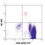
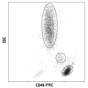
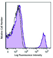
Human peripheral blood lymphocytes, monocytes, granulocytes ... -
Alexa Fluor® 647 anti-mouse/human CD15 (SSEA-1)

F9 (mouse embryonic carcinoma cell line) stained with MC-480... -
Alexa Fluor® 488 anti-mouse/human CD15 (SSEA-1)

F9 (mouse embryonic carcinoma cell line) stained with MC-480... -
FITC anti-mouse/human CD15 (SSEA-1)

F9 cells (mouse embryonic carcinoma cell line) were stained ... -
TotalSeq™-A0076 anti-mouse/human CD15 (SSEA-1)
-
APC anti-mouse/human CD15 (SSEA-1)

Murine embryonic carcinoma cell line F9 was stained with CD1... -
TotalSeq™-C0076 anti-mouse/human CD15 (SSEA-1)
-
TotalSeq™-B0076 anti-mouse/human CD15 (SSEA-1)


Follow Us