


The cell cycle is a series of steps that cells must undergo for replication. Cells can divide in response to stimuli such as growth factors and cytokines, or specific antigens. This response must be tightly regulated, since improper cell proliferation can lead to tumor growth or developmental problems. Study cell division with antibodies for detecting cyclins and cyclin-dependent kinases that control cycle progression, bioactive and stable recombinant proteins that stimulate or suppress cell division, and specialized dyes that can track cell proliferation.
Loading...

Cell Cycle Phases
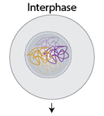 |
Interphase – During interphase, which is comprised of G1, S, and G2 phases, the cell prepares to undergo division by replicating its genome and organelles. DNA replication is performed by DNA polymerase, with the help of factors like the PCNA clamp and topoisomerase-II α (TOP2A). Progression through checkpoints during interphase is regulated by cyclin-dependent kinases (CDKs), which are activated when bound by specific cyclins (see table below). |
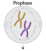 |
Prophase – Prophase is the first step of mitosis, a type of cell division that produces two daughter cells with identical genomes. During prophase, DNA condenses to form tightly packed chromosomes, the nuclear membrane begins to break down, and the mitotic spindle takes shape. Detect histone modifications that regulate DNA compaction |
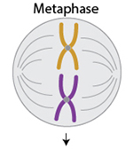 |
Metaphase – Chromosomes are lined along the middle of the cell, and microtubules attach to chromosome kinetochores. Proteins like TRF2 help protect chromosome structures and telomere ends as mitosis progresses. |
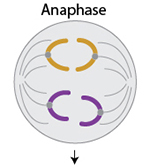 |
Anaphase – Microtubules pull each chromatid to opposite poles of the cell, so that each daughter cell will have a copy of the genome. TPX2 is a protein critical for microtubule growth and mitotic spindle function. View antibodies for tubulin, a major component of microtubules |
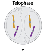 |
Telophase – Each new daughter cell begins to recover from replication by loosening DNA compaction, reforming nuclear membranes, and breaking down the mitotic spindle. |
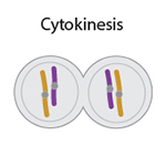 |
Cytokinesis – Towards the end of mitosis, the parent cell physically splits into two cells, a process called cytokinesis. This is regulated by factors like PRC1, a microtubule-associated protein. |
Cyclins and CDKs. The roles of major cyclin-dependent kinases (CDKs) when they are activated by each cyclin.
| Cyclin | Cyclin-dependent kinase | Function |
|---|---|---|
|
CDK4, CDK6 |
Regulates progress through G1 |
|
|
Cyclin E |
Initiates G1 to S transition, and needed to clear restriction point |
|
|
Regulates progress through S |
||
|
CDK1 (Cdc2) |
Drives entry into M |
|
|
N/A |
Regulates neuronal development |
Using DNA Dyes to Assess the Cell Cycle
One of the most common ways to visualize the cell cycle is by using DNA dyes that bind stoichiometrically to dsDNA. Cell permeant dyes like Cytophase™ Violet and DRAQ5™ can measure the cell cycle in live cells while membrane impermeant dyes like Propidium Iodide, DRAQ7™, and Helix NP™ can only assess cell cycle status in cells that have been fixed or permeabilized.
To more accurately measure the number of cells in S phase, BrdU can be used in addition to a DNA dye. BrdU is incorporated into DNA during S-phase of the cell cycle as a thymidine substitute. Cells can be pulse-labeled with BrdU and any cells in S-phase during that time interval will be positive when stained with an anti-BrdU antibody.
CytoPhase™ Violet is a cell-permeant DNA-binding dye that can be excited by either the UV (355 nm or 375 nm) or violet laser (405 nm). It can be used for flow cytometric analysis of the cell cycle in live or fixed cells, and can be used concurrently with antibody staining procedures.
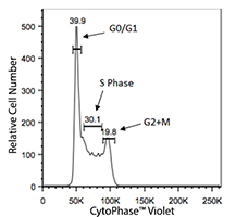
Live Ramos cells stained with 5 µM CytoPhase™ Violet. A typical cell cycle profile will contain two distinct peaks representing cells in G0/G1 and G2/M. Cells that fall between these peaks are in S-phase.
Phase-Flow™ kits are designed for streamlined analysis of BrdU incorporation by flow cytometry. The kits provide BrdU pulsing solution, anti-BrdU antibody, all necessary buffers, and DNA dyes DAPI and 7-AAD.

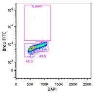
Ramos cells loaded with BrdU for 1.5 hours (left) or as no load controls (right) stained using the Phase-Flow™ FITC BrdU Kit and DAPI.
View products for:
BrdU | Propidium Iodide | Cytophase™ Violet | DRAQ5 and DRAQ7™ | Helix NP™
Growth Regulation
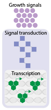 The initiation of cell replication begins with extracellular signals. For human and animal cells, this is usually triggered by growth factors or cytokines/chemokines.
View growth factors or cytokines/chemokines.
When extracellular signals are received, signal transduction pathways are initiated by proteins like kinases and phosphatases.
View products for kinases/phosphatases that regulate the cell cycle.
Signaling pathways ultimately lead to the activation of transcription factors that regulate genes involved in the cell cycle.
View products for transcription factors involved in the cell cycle.
Cell cycle inhibition – Dysregulation of the cell cycle can result in uncontrolled proliferation of cells, which will cause tumor growth and cancer. p53 is a major tumor-suppressor that can induce arrest of the cell cycle at the G1 phase and cause apoptosis. Another critical inhibitor of the cell cycle is CDKN2A, which negatively regulates CDK4 and CDK6.
Tracking cell proliferation
Proliferation of cells can be measured using probes that are retained by daughter cells, nuclear markers that are only expressed by cycling cells, or assays that assess metabolic activity. The table below summarizes kits and reagents for tracking cell proliferation.
| Reagent or Kit | Description |
|---|---|
| CFSE | Retained in daughter cells following cell division, can be used for ex vivo and in vivo cell proliferation assays. Its peak excitation and emission wavelengths are 492 nm and 517 nm, respectively. |
| Tag-it™ Violet | Retained in daughter cells following cell division, can be used for ex vivo and in vivo cell proliferation assays. Its peak excitation and emission wavelengths are 395 nm and 455 nm, respectively. |
| BrdU | Incorporated into newly synthesized DNA, and can be detected with anti-BrdU antibodies. |
| Phase-Flow™ BrdU kits | Complete set of reagents for analyzing BrdU incorporation by flow cytometry. Kit includes BrdU pulsing solution, anti-BrdU antibody, all necessary buffers, and DNA dyes DAPI and 7-AAD. |
| Ki-67 | Nuclear protein expressed only in cycling cells, and not found in quiescent or senescent (G0) cells. |
| Deep Blue Cell Viability™ | Measures resazurin reduction to determine cell metabolic activity and rate. |
| LDH-Cytox Assay™ Kit | Measures tetrazolium salt reduction resulting from lactate dehydrogenase (LDH) activity. Since LDH is released from damaged cells, this assay can be used to determine cytotoxicity. |
ProductsHere


Follow Us