 Login / Register


- Clone
- 100 (See other available formats)
- Regulatory Status
- RUO
- Other Names
- B-Cell CLL/Lymphoma 2 (Bcl-2), Protein Phosphatase 1, Regulatory Subunit 50
- Isotype
- Mouse IgG1
- Ave. Rating
- Submit a Review
- Product Citations
- publications

-

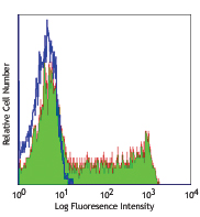
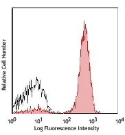
Human peripheral blood lymphocytes were treated with Fixation Buffer (Cat. No. 420801) and Permeabilization Wash Buffer (Cat. No. 421002), then stained with Bcl-2 (clone 100) Alexa Fluor® 647 (filled histogram) or mouse IgG1, κ Alexa Fluor® 647 isotype control (open histogram). -

Confocal image of human lymph node sample acquired using the IBEX method of highly multiplexed antibody-based imaging: BCL2 (cyan) in Cycle 1, CD3 (purple) in Cycle 4. Tissues were prepared using ~1% (vol/vol) formaldehyde and a detergent. Following fixation, samples are immersed in 30% (wt/vol) sucrose for cryoprotection. Images are courtesy of Drs. Andrea J. Radtke and Ronald N. Germain of the Center for Advanced Tissue Imaging (CAT-I) in the National Institute of Allergy and Infectious Diseases (NIAID, NIH).
| Cat # | Size | Price | Quantity Check Availability | Save | ||
|---|---|---|---|---|---|---|
| 658705 | 25 tests | 100€ | ||||
| 658706 | 100 tests | 249€ | ||||
Bcl-2 is a conserved anti-apoptotic protein that plays important roles in normal immunity. It forms homodimers or heterodimers with other Bcl-2 family members. This protein regulates apoptosis through controlling mitochondrial fusion and fission. Phosphorylation of Bcl-2 has been shown to enhance activity to allow response to extracellular growth-factor-mediated signals. Bcl-2 is also regulated by IRES, miR-15a, miR-16-1 and RNA-BP nucleolin. Chromosome 18q21.3 translocation and overexpression of Bcl-2 is frequently observed in follicular lymphomas and some diffuse large B-cell lymphomas.
Product DetailsProduct Details
- Verified Reactivity
- Human
- Antibody Type
- Monoclonal
- Host Species
- Mouse
- Immunogen
- Synthetic peptide of amino acids 41-54 of human bcl-2 oncoprotein
- Formulation
- Phosphate-buffered solution, pH 7.2, containing 0.09% sodium azide and BSA (origin USA)
- Preparation
- The antibody was purified by affinity chromatography and conjugated with Alexa Fluor® 647 under optimal conditions.
- Concentration
- Lot-specific (to obtain lot-specific concentration and expiration, please enter the lot number in our Certificate of Analysis online tool.)
- Storage & Handling
- The antibody solution should be stored undiluted between 2°C and 8°C, and protected from prolonged exposure to light. Do not freeze.
- Application
-
ICFC - Quality tested
SB - Reported in the literature, not verified in house - Recommended Usage
-
Each lot of this antibody is quality control tested by intracellular immunofluorescent staining with flow cytometric analysis. For flow cytometric staining, the suggested use of this reagent is 5 µl per million cells in 100 µl staining volume or 5 µl per 100 µl of whole blood.
* Alexa Fluor® 647 has a maximum emission of 668 nm when it is excited at 633 nm / 635 nm.
Alexa Fluor® and Pacific Blue™ are trademarks of Life Technologies Corporation.
View full statement regarding label licenses - Excitation Laser
-
Red Laser (633 nm)
- Additional Product Notes
-
Iterative Bleaching Extended multi-pleXity (IBEX) is a fluorescent imaging technique capable of highly-multiplexed spatial analysis. The method relies on cyclical bleaching of panels of fluorescent antibodies in order to image and analyze many markers over multiple cycles of staining, imaging, and, bleaching. It is a community-developed open-access method developed by the Center for Advanced Tissue Imaging (CAT-I) in the National Institute of Allergy and Infectious Diseases (NIAID, NIH).
- Application References
- Product Citations
-
- RRID
-
AB_2563279 (BioLegend Cat. No. 658705)
AB_2563280 (BioLegend Cat. No. 658706)
Antigen Details
- Structure
- Two isoforms, 239 or 205 amino acids in length, with a predicted molecular weight of 26 kD or 22 kD and four ‘BH' domains.
- Distribution
-
Outer mitochondrial membrane, nuclear membrane, endoplasmic reticulum membrane.
- Function
- Regulates apoptosis through controlling mitochondrial fusion and fission.
- Interaction
- BAX, BAD, BAK, Bcl-X(L),EI24,APAF1, BBC3, BCL2L1, BNIPL, MRPL41, TP53BP2, FKBP8 BAG1, RAF1, EGLN3, G0S2, and BOP.
- Cell Type
- B cells
- Biology Area
- Apoptosis/Tumor Suppressors/Cell Death, Cell Biology, Cell Cycle/DNA Replication, Chromatin Remodeling/Epigenetics, Immunology, Mitochondrial Function, Neuroscience, Signal Transduction, Transcription Factors, Ubiquitin/Protein Degradation
- Antigen References
-
1. Lin P, et al. 2013. Curr. Hematol. Malig. Rep. 8:243.
2. Tomita N. 2011. J. Clin. Exp. Hematop. 51:7.
3. Kelly PN, et al. 2011. Cell Death Differ. 18:1414.
4. Willimott S, et al. 2010. Biochem. Soc. Trans. 38:1571.
5. Rolland SG, et al. 2010. Curr. Opin. Cell Biol. 22:852. - Gene ID
- 596 View all products for this Gene ID
- UniProt
- View information about Bcl-2 on UniProt.org
Related Pages & Pathways
Pathways


Related FAQs
- If an antibody clone has been previously successfully used in IBEX in one fluorescent format, will other antibody formats work as well?
-
It’s likely that other fluorophore conjugates to the same antibody clone will also be compatible with IBEX using the same sample fixation procedure. Ultimately a directly conjugated antibody’s utility in fluorescent imaging and IBEX may be specific to the sample and microscope being used in the experiment. Some antibody clone conjugates may perform better than others due to performance differences in non-specific binding, fluorophore brightness, and other biochemical properties unique to that conjugate.
- Will antibodies my lab is already using for fluorescent or chromogenic IHC work in IBEX?
-
Fundamentally, IBEX as a technique that works much in the same way as single antibody panels or single marker IF/IHC. If you’re already successfully using an antibody clone on a sample of interest, it is likely that clone will have utility in IBEX. It is expected some optimization and testing of different antibody fluorophore conjugates will be required to find a suitable format; however, legacy microscopy techniques like chromogenic IHC on fixed or frozen tissue is an excellent place to start looking for useful antibodies.
- Are other fluorophores compatible with IBEX?
-
Over 18 fluorescent formats have been screened for use in IBEX, however, it is likely that other fluorophores are able to be rapidly bleached in IBEX. If a fluorophore format is already suitable for your imaging platform it can be tested for compatibility in IBEX.
- The same antibody works in one tissue type but not another. What is happening?
-
Differences in tissue properties may impact both the ability of an antibody to bind its target specifically and impact the ability of a specific fluorophore conjugate to overcome the background fluorescent signal in a given tissue. Secondary stains, as well as testing multiple fluorescent conjugates of the same clone, may help to troubleshoot challenging targets or tissues. Using a reference control tissue may also give confidence in the specificity of your staining.
- How can I be sure the staining I’m seeing in my tissue is real?
-
In general, best practices for validating an antibody in traditional chromogenic or fluorescent IHC are applicable to IBEX. Please reference the Nature Methods review on antibody based multiplexed imaging for resources on validating antibodies for IBEX.
Other Formats
View All Bcl-2 Reagents Request Custom Conjugation| Description | Clone | Applications |
|---|---|---|
| Purified anti-Bcl-2 | 100 | WB,IHC-F,IHC-P |
| Alexa Fluor® 647 anti-Bcl-2 | 100 | ICFC,SB |
| Alexa Fluor® 488 anti-Bcl-2 | 100 | ICFC |
| PE anti-Bcl-2 | 100 | ICFC |
| Brilliant Violet 421™ anti-Bcl-2 | 100 | ICFC |
| TotalSeq™-Bn1180 anti-Bcl-2 | 100 | SB |
| TotalSeq™-B1180 anti-Bcl-2 | 100 | ICPG |
Customers Also Purchased
Compare Data Across All Formats
This data display is provided for general comparisons between formats.
Your actual data may vary due to variations in samples, target cells, instruments and their settings, staining conditions, and other factors.
If you need assistance with selecting the best format contact our expert technical support team.
-
Purified anti-Bcl-2

Western blot analysis of extracts from THP-1(lane 1), PBMC(l... 
IHC staining using purified anti-Bcl-2 antibody (clone 100) ... -
Alexa Fluor® 647 anti-Bcl-2

Human peripheral blood lymphocytes were treated with Fixatio... 
Confocal image of human lymph node sample acquired using the... -
Alexa Fluor® 488 anti-Bcl-2

Human peripheral blood lymphocytes were treated with Fixatio... -
PE anti-Bcl-2

Human peripheral blood lymphocytes were treated with Fixatio... -
Brilliant Violet 421™ anti-Bcl-2

Human peripheral blood lymphocytes were treated with Fixatio... -
TotalSeq™-Bn1180 anti-Bcl-2
-
TotalSeq™-B1180 anti-Bcl-2


Follow Us