 Login / Register


- Clone
- W16131A (See other available formats)
- Regulatory Status
- RUO
- Other Names
- Keratin, type I cytoskeletal 17, keratin-17 (K-17), cytokeratin-17 (CK-17), KRT17, CK17, K17
- Isotype
- Rat IgG2b, κ
- Ave. Rating
- Submit a Review
- Product Citations
- publications

-

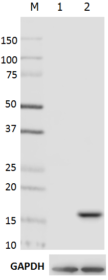
Total cell lysates (15µg protein) from A549, A431 and NIH3T3 cells were resolved by 4-20% Tris-glycine gel electrophoresis, transferred to nitrocellulose, and probed with 0.5µg/mL purified anti- Cytokeratin 17 (clone W16131A) antibody, competitor’s antibody used at manufactures recommended concentration (upper) or loading control anti-GAPDH (poly6314) antibody (lower). Proteins were visualized by chemiluminescence detection using a goat anti-rat-IgG secondary antibody conjugated to HRP for anti- Cytokeratin 17 antibody, and a donkey anti-rabbit IgG Antibody conjugated to HRP for competitor’s antibody and anti-GAPDH antibody. Lane M: Molecular Weight ladder. -

A431 cells were fixed with 4% paraformaldehyde (PFA) for fifteen minutes, permeabilized with 0.5% Triton X-100 for three minutes, and blocked with 5% FBS for 60 minutes. Then the cells were intracellularly stained with 2 µg/ml rat IgG2b isotype control (top left), anti-Cytokeratin 17 (clone W16131A, top right), or competitor’s antibody used at manufactures recommended concentration (bottom left) overnight at 4°C followed by Alexa Fluor® 594 (red) conjugated goat anti-rat IgG for anti- Cytokeratin 17 antibody, Alexa Fluor® 594 (red) conjugated donkey anti-rabbit IgG for competitor’s antibody for one hour at room temperature. Nuclei were counterstained with DAPI (blue). The image was captured with a 60X objective. -

IHC staining using purified anti-Cytokeratin 17 (clone W16131A) on formalin-fixed paraffin-embedded human bladder tissue. Following antigen retrieval using 1X Tris-Buffered Saline (final concentration 0.05M) with Tween-20 (Cat. No. 925501), the tissue was incubated with (B) or without (A) 10 μg/mL of antibody overnight at 4°C, followed by incubation with 2.5 μg/mL of Alexa Fluor® 647 goat anti-rat IgG (Cat. No. 405416) for one hour at room temperature. Nuclei were counterstained with DAPI (blue) (Cat. No. 422801), and the slide was mounted with ProLong™ Gold Antifade Mountant. The image was captured with a 10X objective. Scalebar = 50 μM -

IHC staining using purified anti-Cytokeratin 17 (clone W16131A) on formalin-fixed paraffin-embedded human bladder tissue. Following antigen retrieval using 1X Tris-Buffered Saline (final concentration 0.05M) with Tween-20 (Cat. No. 925501), the tissue was incubated with (B) or without (A) 10 μg/mL of antibody overnight at 4°C, followed by incubation with 2.5 μg/mL of Alexa Fluor® 647 goat anti-rat IgG (Cat. No. 405416) for one hour at room temperature. Nuclei were counterstained with DAPI (blue) (Cat. No. 422801), and the slide was mounted with ProLong™ Gold Antifade Mountant. The image was captured with a 40X objective. Scalebar = 50
| Cat # | Size | Price | Quantity Check Availability | Save | ||
|---|---|---|---|---|---|---|
| 697202 | 100 µg | 221€ | ||||
Cytokeratin 17 (CK-17), also known as KRT17 or keratin-17 (K-17), is a type I intermediate filament protein. KRT expression is normally restricted to ectoderm-derived epithelial appendages such as thymus, glands, skin, hair, tooth, nail, and the endocervical mucosa. Cytokeratin-17 participates in promoting protein synthesis and cell growth through recruiting stratifin (SFN) to the cytoplasm and stimulating the Akt/mTOR pathway. Cytokeratin-17 was found to regulate chemokine expression, including the CXCR3 ligands CXCL9, CXCL10, and CXCL11. Cytokeratin 17 expression can be upregulated in response to chemical stimulation, wounding, and UV irradiation. Increased expression of Cytokeratin 17 is associated with lesion progression and poor prognosis in epithelial carcinoma. Cytokeratin 17 deficient mice show a delay in wound healing and attenuated tumorigenesis.
Product DetailsProduct Details
- Verified Reactivity
- Human
- Antibody Type
- Monoclonal
- Host Species
- Rat
- Immunogen
- Human Cytokeratin 17 peptide (415-432 a.a.) conjugated to KLH
- Formulation
- Phosphate-buffered solution, pH 7.2, containing 0.09% sodium azide.
- Preparation
- The antibody was purified by affinity chromatography.
- Concentration
- 0.5 mg/ml
- Storage & Handling
- The antibody solution should be stored undiluted between 2°C and 8°C.
- Application
-
WB - Quality tested
ICC, IHC-P - Verified - Recommended Usage
-
Each lot of this antibody is quality control tested by Western blotting. For Western blotting, the suggested use of this reagent is 0.05 - 0.5 µg per ml. For immunocytochemistry, a concentration range of 0.2 - 2.0 µg per ml is recommended. For immunohistochemistry on formalin-fixed paraffin-embedded tissue sections, a concentration of 10.0 µg/mL is suggested. It is recommended that the reagent be titrated for optimal performance for each application.
- Application Notes
-
This clone does not cross-react with mouse (in-house tested).
As per the Human Protein Atlas RNA level dataset, A549 cell is expected to have a low level of Cytokeratin 17 expression. - RRID
-
AB_2687136 (BioLegend Cat. No. 697202)
Antigen Details
- Structure
- 432 amino acids with a predicted molecular weight of 48 kD. Belongs to the type-I intermediate filament family.
- Distribution
-
Cytoplasm
- Function
- Cytokeratin 17 is a type I intermediate filament protein and is involved in promoting cell growth, chemokine production, and tumorigenesis.
- Interaction
- Forms a heterodimer with a type I or a type II keratin. Interacts with TRADD and SFN.
- Biology Area
- Cancer Biomarkers, Cell Biology, Cell Motility/Cytoskeleton/Structure, Neuroscience, Neuroscience Cell Markers
- Molecular Family
- Intermediate Filaments
- Antigen References
-
1. Hobbs RP, et al. 2016. Oncogene. 35:5653.
2. Escobar-Hoyos LF, et al. 2015. Cancer Res. 75:3650.
3. Chung BM, et al. 2015. J. Cell. Biol. 208:613.
4. Sankar S, et al. 2013. Mol. Cell. Biol. 33:4448.
5. Jin L, et al. 2014. Med. Res. Rev. 34:438.
6. Depianto D, et al. 2010. Nat. Genet. 42:910. - Gene ID
- 3872 View all products for this Gene ID
- UniProt
- View information about Cytokeratin 17 on UniProt.org

Related FAQs
Other Formats
View All Cytokeratin 17 Reagents Request Custom Conjugation| Description | Clone | Applications |
|---|---|---|
| Purified anti-Cytokeratin 17 | W16131A | WB,ICC,IHC-P |
| Alexa Fluor® 594 anti-Cytokeratin 17 | W16131A | ICC |
| Alexa Fluor® 647 anti-Cytokeratin 17 | W16131A | ICC |
| Alexa Fluor® 488 anti-Cytokeratin 17 | W16131A | ICC |
Customers Also Purchased
Compare Data Across All Formats
This data display is provided for general comparisons between formats.
Your actual data may vary due to variations in samples, target cells, instruments and their settings, staining conditions, and other factors.
If you need assistance with selecting the best format contact our expert technical support team.
-
Purified anti-Cytokeratin 17

Total cell lysates (15µg protein) from A549, A431 and NIH3T3... 
A431 cells were fixed with 4% paraformaldehyde (PFA) for fi... 
IHC staining using purified anti-Cytokeratin 17 (clone W1613... 
IHC staining using purified anti-Cytokeratin 17 (clone W1613... -
Alexa Fluor® 594 anti-Cytokeratin 17

A431 cells were fixed with 4% paraformaldehyde (PFA) for 10 ... -
Alexa Fluor® 647 anti-Cytokeratin 17

HeLa cells were fixed with 4% paraformaldehyde for 10 minute... -
Alexa Fluor® 488 anti-Cytokeratin 17

HeLa cells were fixed with 4% paraformaldehyde for 10 minute...


Follow Us