 Login / Register


- Clone
- W16155A (See other available formats)
- Regulatory Status
- RUO
- Other Names
- Keratin type II cytoskeletal 7, Cytokeratin-7, CK-7, Keratin-7, K7, Type-II keratin Kb7, Sarcolectin, SCL, Cytokeratin 7, Keratin 7
- Isotype
- Rat IgG2a, κ
- Ave. Rating
- Submit a Review
- Product Citations
- publications

-

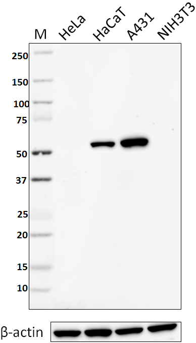
Total cell lysate (15ug protein) from HaCaT, A549, MCF-7 (negative control) and NIH3T3 cells were resolved by electrophoresis (4-12% Bis-Tris gel), transferred to nitrocellulose, and probed with 1:250 (2 µg/mL) diluted purified anti-KRT7 antibody (clone W16155A) or competitor’s antibody used at manufactures recommended concentration (upper). Proteins were visualized by chemiluminescence detection using 1:3000 diluted HRP rat-IgG secondary antibody conjugated to HRP (clone Poly4054) for clone W16155A or 1:3000 diluted donkey anti-rabbit IgG Antibody conjugated to HRP (clone Poly4064) for competitor’s antibody (upper). 1:2000 dilution of Direct-Blot™ HRP anti-β-actin antibody (clone 2F1-1) was used as a loading control (lower). Lane M: MW ladder, M* indicates longer exposure. -

HaCaT cells were fixed with 4% paraformaldehyde (PFA) for 15 minutes, permeabilized with 0.5% Triton X-100 for 3 minutes, and blocked with 5% FBS for 60 minutes. Then the cells were intracellularly stained with 1:250 diluted (2 µg/ml) anti-KRT7 (clone W16155A) antibody overnight at 4 degrees followed by 1:250 diluted (2 µg/ml) Alexa Fluor® 594 (red) conjugated goat anti-rat IgG (clone Poly4054) antibody for one hour at room temperature. Nuclei were counterstained with DAPI (blue). The image was captured with a 60X objective. -

HaCaT cells were fixed with 4% paraformaldehyde (PFA) for 15 minutes, permeabilized with 0.5% Triton X-100 for 3 minutes, and blocked with 5% FBS for 60 minutes. Then the cells were intracellularly stained with 1:250 diluted (2 μg/ml) rat IgG2b, κ isotype control (clone RTK4530) overnight at 4 degrees followed by 1:250 diluted (2 μg/ml) Alexa Fluor® 594 (red) conjugated goat anti-rat IgG (clone Poly4054) antibody for one hour at room temperature. Nuclei were counterstained with DAPI (blue). The image was captured with a 60X objective.
| Cat # | Size | Price | Quantity Check Availability | Save | ||
|---|---|---|---|---|---|---|
| 601601 | 25 µg | 81€ | ||||
| 601602 | 100 µg | 203€ | ||||
Keratin 7 (KRT7) belongs to type II keratin and is expressed in the epithelia lining the cavities of internal organs, gland ducts and blood vessels. The function of KRT7 is still not clear but it has been suggested involved in the translational regulation of the human papillomavirus type 16 E7 mRNA (HPV16 E7). In cancer, the expression of KRT7 is observed in many types of carcinoma and is associated with neoplasm progression. The diverse and unique expression pattern of KRT7 is often used as a diagnosis indicator in epithelial origin carcinoma. For example, aberrant KRT7 expression is observed in triple negative breast cancer. In bladder cancer, loss of KRT7 results in promoting proliferation of the bladder urothelium.
Product DetailsProduct Details
- Verified Reactivity
- Human
- Antibody Type
- Monoclonal
- Host Species
- Rat
- Immunogen
- Human KRT7 peptide (446-462) conjugated to KLH.
- Formulation
- Phosphate-buffered solution, pH 7.2, containing 0.09% sodium azide.
- Preparation
- The antibody was purified by affinity chromatography.
- Concentration
- 0.5 mg/ml
- Storage & Handling
- The antibody solution should be stored undiluted between 2°C and 8°C.
- Application
-
WB - Quality tested
ICC - Verified - Recommended Usage
-
Each lot of this antibody is quality control tested by Western blotting. For Western blotting, the suggested use of this reagent is 0.2 - 2.0 µg per ml (1:250 - 1:2500 dilution). For immunocytochemistry, the recommended usage is 0.2 - 2.0 µg per mL (1:250 - 1:2500 dilution). It is recommended that the reagent be titrated for optimal performance for each application.
- Application Notes
-
This antibody does not react with mouse (in-house tested). Additional reported applications (for the relevant formats) include: spatial biology (IBEX)1,2.
- Application References
- Product Citations
-
- RRID
-
AB_2715904 (BioLegend Cat. No. 601601)
AB_2715905 (BioLegend Cat. No. 601602)
Antigen Details
- Structure
- Type II cytokeratin. 469 amino acids with a predicted molecular weight of 51 kD.
- Distribution
-
Cytoplasm.
- Function
- KRT7 is a type II keratin, involved in the translational regulation of the human papillomavirus type 16 E7 mRNA.
- Interaction
- KRT7 interacts with EIF3S10, HPV16 E7, and GPER1.
- Biology Area
- Cell Biology, Cell Motility/Cytoskeleton/Structure, Neuroscience, Neuroscience Cell Markers
- Molecular Family
- Intermediate Filaments
- Antigen References
-
1. Tajima Y, et al. 2017. Sci Rep. 7:40684.
2. Huang B, et al. 2016. Oncogene. 35:4927.
3. Szponar A, et al. 2012. Virchows Arch. 460:423.
4. Sano M, et al. 2010. Int J Oncol. 36:321. - Gene ID
- 3855 View all products for this Gene ID
- UniProt
- View information about Cytokeratin 7 on UniProt.org

Related FAQs
Other Formats
View All Cytokeratin 7 Reagents Request Custom Conjugation| Description | Clone | Applications |
|---|---|---|
| Purified anti-human Cytokeratin 7 | W16155A | WB,ICC |
| Alexa Fluor® 594 anti-human Cytokeratin 7 | W16155A | ICC |
| Alexa Fluor® 488 anti-human Cytokeratin 7 | W16155A | ICC,SB |
| Alexa Fluor® 647 anti-human Cytokeratin 7 | W16155A | ICC |
Customers Also Purchased
Compare Data Across All Formats
This data display is provided for general comparisons between formats.
Your actual data may vary due to variations in samples, target cells, instruments and their settings, staining conditions, and other factors.
If you need assistance with selecting the best format contact our expert technical support team.
-
Purified anti-human Cytokeratin 7

Total cell lysate (15ug protein) from HaCaT, A549, MCF-7 (ne... 
HaCaT cells were fixed with 4% paraformaldehyde (PFA) for 1... 
HaCaT cells were fixed with 4% paraformaldehyde (PFA) for 15... -
Alexa Fluor® 594 anti-human Cytokeratin 7

HaCaT cells were fixed with 4% paraformaldehyde (PFA) for 1... 
HaCaT cells were fixed with 4% paraformaldehyde (PFA) for 1... -
Alexa Fluor® 488 anti-human Cytokeratin 7

HaCaT cells were fixed with 4% paraformaldehyde (PFA) for 10... 
Confocal image of human liver sample acquired using the IBEX... -
Alexa Fluor® 647 anti-human Cytokeratin 7

HaCaT cells were fixed with 4% paraformaldehyde (PFA) for 10...


Follow Us