 Login / Register


- Clone
- RMP1-30 (See other available formats)
- Regulatory Status
- RUO
- Other Names
- Programmed Death-1, CD279, PDCD1
- Isotype
- Rat IgG2b, κ
- Ave. Rating
- Submit a Review
- Product Citations
- publications

-

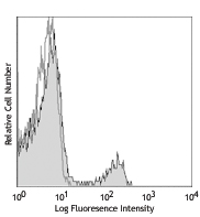
Con A-stimulated (day-3) Balb/c mouse splenocytes stained with RMP1-30 PE/Cyanine7
| Cat # | Size | Price | Quantity Check Availability | Save | ||
|---|---|---|---|---|---|---|
| 109109 | 25 µg | 88€ | ||||
| 109110 | 100 µg | 234€ | ||||
CD279 is a 50-55 kD immunoglobulin superfamily member also known as programmed death-1 (PD-1). PD-1 is expressed on a subset of CD4-CD8- thymocytes and on activated T and B cells. PD-1 is thought to be involved in lymphocyte clonal selection and peripheral tolerance. The PD-1 ligands, PD-L1 (also known as B7-H1) and PD-L2 (B7-DC), are members of the B7 immunoglobulin superfamily.
Product DetailsProduct Details
- Verified Reactivity
- Mouse
- Antibody Type
- Monoclonal
- Host Species
- Rat
- Immunogen
- Mouse PD-1 transfected BHK cells
- Formulation
- Phosphate-buffered solution, pH 7.2, containing 0.09% sodium azide.
- Preparation
- The antibody was purified by affinity chromatography, and conjugated with PE/Cyanine7 under optimal conditions.
- Concentration
- 0.2 mg/ml
- Storage & Handling
- The CD279 antibody solution should be stored undiluted between 2°C and 8°C, and protected from prolonged exposure to light. Do not freeze.
- Application
-
FC - Quality tested
- Recommended Usage
-
Each lot of this antibody is quality control tested by immunofluorescent staining with flow cytometric analysis. For flow cytometric staining, the suggested use of this reagent is =1.0 µg per million cells in 100 µl volume. It is recommended that the reagent be titrated for optimal performance for each application.
- Excitation Laser
-
Blue Laser (488 nm)
Green Laser (532 nm)/Yellow-Green Laser (561 nm)
- Application Notes
-
The RMP1-30 antibody does not block the binding of PD-1 to B7-H1 and B7-DC1.
- Additional Product Notes
- BioLegend is in the process of converting the name PE/Cy7 to PE/Cyanine7. The dye molecule remains the same, so you should expect the same quality and performance from our PE/Cyanine7 products. Please contact Technical Service if you have any questions.
-
Application References
(PubMed link indicates BioLegend citation) - Product Citations
-
- RRID
-
AB_572016 (BioLegend Cat. No. 109109)
AB_572017 (BioLegend Cat. No. 109110)
Antigen Details
- Structure
- Ig superfamily, 50-55 kD
- Distribution
-
Subset of double negative thymocytes, activated T and B cells
- Function
- Lymphocyte clonal selection, peripheral tolerance
- Ligand/Receptor
- PD-L1 (B7-H1), PD-L2
- Cell Type
- B cells, T cells, Thymocytes, Tregs
- Biology Area
- Apoptosis/Tumor Suppressors/Cell Death, Cancer Biomarkers, Cell Biology, Immunology, Inhibitory Molecules
- Molecular Family
- CD Molecules, Immune Checkpoint Receptors
- Antigen References
-
1. Barclay A, et al. 1997. The Leukocyte Antigen FactsBook Academic Press.
2. Agata Y, et al. 1996. Int. Immunol. 8:765.
3. Nishimura H, et al. 2001. Science 291:319.
4. Ishida Y, et al. 1992. EMBO J. 11:3887. - Gene ID
- 18566 View all products for this Gene ID
- Specificity (DOES NOT SHOW ON TDS):
- CD279
- Specificity Alt (DOES NOT SHOW ON TDS):
- CD279
- App Abbreviation (DOES NOT SHOW ON TDS):
- FC
- UniProt
- View information about CD279 on UniProt.org
Related Pages & Pathways
Pathways


Related FAQs
Other Formats
View All CD279 Reagents Request Custom Conjugation| Description | Clone | Applications |
|---|---|---|
| PE anti-mouse CD279 (PD-1) | RMP1-30 | FC |
| Purified anti-mouse CD279 (PD-1) | RMP1-30 | FC,IHC-F |
| Biotin anti-mouse CD279 (PD-1) | RMP1-30 | FC |
| LEAF™ Purified anti-mouse CD279 (PD-1) | RMP1-30 | FC, FA |
| PE/Cyanine7 anti-mouse CD279 (PD-1) | RMP1-30 | FC |
| APC anti-mouse CD279 (PD-1) | RMP1-30 | FC |
| Purified anti-mouse CD279 (PD-1) (Maxpar® Ready) | RMP1-30 | FC,CyTOF® |
| PE/Dazzle™ 594 anti-mouse CD279 (PD-1) | RMP1-30 | FC |
| Alexa Fluor® 647 anti-mouse CD279 (PD-1) | RMP1-30 | FC |
| PerCP/Cyanine5.5 anti-mouse CD279 (PD-1) | RMP1-30 | FC |
| Brilliant Violet 421™ anti-mouse CD279 (PD-1) | RMP1-30 | FC |
| TotalSeq™-A0004 anti-mouse CD279 (PD-1) | RMP1-30 | PG |
| TotalSeq™-B0004 anti-mouse CD279 (PD-1) | RMP1-30 | PG |
| TotalSeq™-C0004 anti-mouse CD279 (PD-1) | RMP1-30 | PG |
| KIRAVIA Blue 520™ anti-mouse CD279 (PD-1) | RMP1-30 | FC |
Customers Also Purchased
Compare Data Across All Formats
This data display is provided for general comparisons between formats.
Your actual data may vary due to variations in samples, target cells, instruments and their settings, staining conditions, and other factors.
If you need assistance with selecting the best format contact our expert technical support team.
-
PE anti-mouse CD279 (PD-1)

Con A (3-day) activated C57BL/6 mouse splenocytes stained wi... -
Purified anti-mouse CD279 (PD-1)
Con A (3-day) activated C57BL/6 mouse splenocytes stained wi... 
Fresh, frozen mouse spleen was stained with purified CD279 c... -
Biotin anti-mouse CD279 (PD-1)
Con A (3-day) activated C57BL/6 mouse splenocytes stained wi... -
LEAF™ Purified anti-mouse CD279 (PD-1)
-
PE/Cyanine7 anti-mouse CD279 (PD-1)

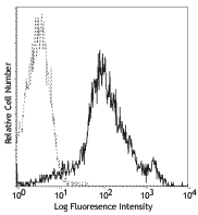
Con A-stimulated (day-3) Balb/c mouse splenocytes stained wi... -
APC anti-mouse CD279 (PD-1)

Con A-stimulated C57BL/6 splenocytes (Day 3) stained with RM... -
Purified anti-mouse CD279 (PD-1) (Maxpar® Ready)

BALB/c mouse splenocytes were incubated for 3 days in media ... -
PE/Dazzle™ 594 anti-mouse CD279 (PD-1)

Con A (three-days) activated C57BL/6 splenocytes were staine... 
-
Alexa Fluor® 647 anti-mouse CD279 (PD-1)

Con A (three-days) activated C57BL/6 splenocytes were staine... 
-
PerCP/Cyanine5.5 anti-mouse CD279 (PD-1)

Con A+IL-2-stimulated C57BL/6 splenocytes (3 days) were stai... -
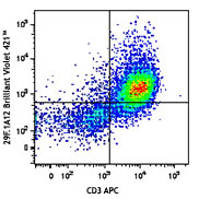
Brilliant Violet 421™ anti-mouse CD279 (PD-1)

Con A (three-days) activated C57BL/6 splenocytes were staine... 
-
TotalSeq™-A0004 anti-mouse CD279 (PD-1)
-
TotalSeq™-B0004 anti-mouse CD279 (PD-1)
-
TotalSeq™-C0004 anti-mouse CD279 (PD-1)
-
KIRAVIA Blue 520™ anti-mouse CD279 (PD-1)

Con A (three-days) activated C57BL/6 splenocytes were staine...


Follow Us