 Login / Register


- Clone
- 17A2 (See other available formats)
- Regulatory Status
- RUO
- Other Names
- T cell antigen receptor complex, T3
- Isotype
- Rat IgG2b, κ
- Ave. Rating
- Submit a Review
- Product Citations
- publications

Watch the video.
-

Paraformaldehyde-fixed (4%), 500 μm-thick mouse spleen section was processed according to the Ce3DTM Tissue Clearing Kit protocol (cat. no. 427701). The section was costained with anti-mouse CD3 Antibody (clone 17A2) Alexa Fluor® 488 at 5 µg/mL (green), anti-mouse IgD Antibody (clone 11-26c.2a) Alexa Fluor® 594 at 5 µg/mL (blue), and anti-mouse CD68 Antibody (clone FA-11) Alexa Fluor® 647 at 5 µg/mL (magenta). The section was then optically cleared and mounted in a sample chamber. The image was captured with a 10X objective using Zeiss 780 confocal microscope and processed by Imaris image analysis software.
Watch the video. -

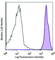
C57BL/6 mouse splenocytes stained with 17A2 Alexa Fluor® 488 -

C57BL/6 mouse frozen spleen section was fixed with 4% paraformaldehyde (PFA) for 10 minutes at room temperature and blocked with 5% FBS for 30 minutes at room temperature. Then, the section was stained with 10 µg/mL CD3 (Clone 17A2) Alexa Fluor® 488 (green) and B220 (Clone RA3-6B2) Brilliant Violet 421™ (blue), overnight at 4°C. The image was captured by 10X objective. -
Formalin-fixed, 300 micron-thick mouse spleen section was blocked, permeabilized and stained overnight with CD3 (clone 17A2) Alexa Fluor® 488 (red), CD21/35 (CR2/CR1)(clone 7E9) Alexa Fluor® 594 (green), and CD45R/B220 (clone RA3-6B2) Alexa Fluor® 647 (blue) all at 5 µg/mL, optically cleared, then analyzed at 215 μm imaging depth on a confocal microscope.

| Cat # | Size | Price | Quantity Check Availability | Save | ||
|---|---|---|---|---|---|---|
| 100212 | 25 µg | 81€ | ||||
| 100210 | 100 µg | 184€ | ||||
CD3, also known as T3, is a member of the Ig superfamily and primarily expressed on T cells, NK-T cells, and at different levels on thymocytes during T cell differentiation. CD3 is composed of CD3ε, δ, γ and ζ chains. It forms a TCR complex by associating with TCR α/β or γ/δ chains. CD3 plays a critical role in TCR signal transduction, T cell activation, and antigen recognition by binding the peptide/MHC antigen complex
Product DetailsProduct Details
- Reactivity
- Mouse
- Antibody Type
- Monoclonal
- Host Species
- Rat
- Immunogen
- γδTCR-positive T-T hybridoma D1
- Formulation
- Phosphate-buffered solution, pH 7.2, containing 0.09% sodium azide.
- Preparation
- The antibody was purified by affinity chromatography and conjugated with Alexa Fluor® 488 under optimal conditions.
- Concentration
- 0.5 mg/mL
- Storage & Handling
- The antibody solution should be stored undiluted between 2°C and 8°C, and protected from prolonged exposure to light. Do not freeze.
- Application
-
FC - Quality tested
IHC-F, 3D IHC - Verified - Recommended Usage
-
Each lot of this antibody is quality control tested by immunofluorescent staining with flow cytometric analysis. For flow cytometric staining, the suggested use of this reagent is ≤1.0 µg per million cells in 100 µL volume. For immunohistochemical staining on frozen tissue sections, the suggested use of this reagent is 5.0 - 10 µg per mL. For 3D immunohistochemistry on formalin-fixed tissues, a concentration of 5.0 µg/mL is suggested. It is recommended that the reagent be titrated for optimal performance for each application.
* Alexa Fluor® 488 has a maximum emission of 519 nm when it is excited at 488 nm.
Alexa Fluor® and Pacific Blue™ are trademarks of Life Technologies Corporation.
View full statement regarding label licenses - Excitation Laser
-
Blue Laser (488 nm)
- Application Notes
-
Additional reported application (for relevant formats) include: spatial biology (IBEX)1,2.
-
Application References
(PubMed link indicates BioLegend citation) - Product Citations
- RRID
-
AB_493530 (BioLegend Cat. No. 100212)
AB_389301 (BioLegend Cat. No. 100210)
Antigen Details
- Structure
- Ig superfamily, CD3/TCR, 20 kD
- Distribution
-
Thymocytes (differentiation dependent), mature T cells, NK-T cells
- Function
- Antigen recognition, TCR signal transduction, T cell activation
- Ligand/Receptor
- Peptide antigen/MHC-complex
- Antigen References
-
1. Barclay A, et al. 1997. The Leukocyte Antigen FactsBook Academic Press.
2. Davis MM. 1990. Annu. Rev. Biochem. 59:475.
3. Weiss A, et al. 1994. Cell 76:263. - Gene ID
- 12502 View all products for this Gene ID
- UniProt
- View information about CD3 on UniProt.org


Customers Also Purchased
Compare Data Across All Formats
This data display is provided for general comparisons between formats.
Your actual data may vary due to variations in samples, target cells, instruments and their settings, staining conditions, and other factors.
If you need assistance with selecting the best format contact our expert technical support team.


Follow Us