 Login / Register


- Clone
- HI100 (See other available formats)
- Regulatory Status
- RUO
- Workshop
- IV N906
- Other Names
- GP180, L-CA, LCA, LY5, T200, PTPRC
- Isotype
- Mouse IgG2b, κ
- Ave. Rating
- Submit a Review
- Product Citations
- publications

-

Human peripheral blood lymphocytes stained with HI100 Alexa Fluor® 700
| Cat # | Size | Price | Quantity Check Availability | Save | ||
|---|---|---|---|---|---|---|
| 304119 | 25 µg | 100€ | ||||
| 304120 | 100 µg | 212€ | ||||
CD45RA is a 205-220 kD single chain type I glycoprotein. It is an exon 4 splice variant of the tyrosine phosphatase CD45. The CD45RA isoform is expressed on resting/naïve T cells, medullary thymocytes, B cells and monocytes. CD45RA enhances both T cell receptor and B cell receptor signaling. CD45 non-covalently associates with lymphocyte phosphatase-associated phosphoprotein (LPAP) on T and B lymphocytes. CD45 has been reported to be associated with several other cell surface antigens including CD1, CD2, CD3, and CD4. CD45 has also been reported to bind galectin-1. CD45 isoform expression can change in response to cytokines.
Product DetailsProduct Details
- Reactivity
- Human
- Antibody Type
- Monoclonal
- Host Species
- Mouse
- Formulation
- Phosphate-buffered solution, pH 7.2, containing 0.09% sodium azide.
- Preparation
- The antibody was purified by affinity chromatography and conjugated with Alexa Fluor® 700 under optimal conditions.
- Concentration
- 0.5 mg/ml
- Storage & Handling
- The antibody solution should be stored undiluted between 2°C and 8°C, and protected from prolonged exposure to light. Do not freeze.
- Application
-
FC - Quality tested
- Recommended Usage
-
Each lot of this antibody is quality control tested by immunofluorescent staining with flow cytometric analysis. The suggested use of this reagent is ≤1.0 µg per million cells in 100 µl volume. It is highly recommended that the reagent be titrated for optimal performance for each application.
* Alexa Fluor® 700 has a maximum emission of 719 nm when it is excited at 633 nm / 635 nm. Prior to using Alexa Fluor® 700 conjugate for flow cytometric analysis, please verify your flow cytometer's capability of exciting and detecting the fluorochrome.
Alexa Fluor® and Pacific Blue™ are trademarks of Life Technologies Corporation.
View full statement regarding label licenses - Excitation Laser
-
Red Laser (633 nm)
- Application Notes
-
Additional reported applications (for relevant formats of this clone) include: inhibition of CD45 functions2, immunohistochemical staining of frozen tissue sections3 and formalin-fixed paraffin-embedded tissue sections4, and immunocytochemistry15,16.
-
Application References
(PubMed link indicates BioLegend citation) -
- Knapp W, et al. 1989. Leucocyte Typing IV. Oxford University Press. New York.
- Yamada T, et al. 2002. J. Biol. Chem. 277:28830. (WB, Block)
- Weninger W, et al. 2003 J. Immunol. 170:4638. (IHC-F)
- Imanguli MM, et al. 2009. Blood. 113:3620 (IHC-P)
- Roque S, et al. 2007. J. Immunol. 178:8028. (FC) PubMed
- Smeltz RB. 2007. J. Immunol. 178:4786. (FC) PubMed
- Palendira U, et al. 2008. Blood (FC) PubMed
- Kuttruff S, et al. 2009. Blood 113:358. (FC) PubMed
- Thakral D, et al. 2008. J. Immunol. 180:7431. (FC) PubMed
- Alanio C, et al. 2010. Blood 115:3718. (FC) PubMed
- Iannello A, et al. 2010. J. Immunol. 184:114. (FC) PubMed
- Yoshino N, et al. 2000. Exp. Anim. (Tokyo) 49:97. (FC)
- Guereau-de-Arellan M, et al. 2011. Brain. 134:3578. PubMed
- Canque B, et al. 2000. Blood 96:3748. (ICC)
- Imanguli MM, et al. 2009. Blood 13:3620. (ICC)
- Stoeckius M, et al. 2017. Nat. Methods. 14:865. (PG)
- Peterson VM, et al. 2017. Nat. Biotechnol. 35:936. (PG)
- Product Citations
- RRID
-
AB_493762 (BioLegend Cat. No. 304119)
AB_493763 (BioLegend Cat. No. 304120)
Antigen Details
- Structure
- Tyrosine phosphatases, type I transmembrane (exon 4 splicing of CD45 gene), 205-220 kD
- Distribution
-
B cells, naïve T cells, monocytes
- Function
- Enhances TCR and BCR signaling
- Ligand/Receptor
- Galectin-1, CD2, CD3, CD4
- Cell Type
- B cells, Monocytes, T cells, Tregs
- Biology Area
- Cell Biology, Immunology, Inhibitory Molecules, Neuroscience, Neuroscience Cell Markers
- Molecular Family
- CD Molecules
- Antigen References
-
1. Thomas M. 1989. Annu. Rev. Immunol. 7:339.
2. Trowbridge I, et al. 1994. Annu. Rev. Immunol.12:85. - Gene ID
- 5788 View all products for this Gene ID
- UniProt
- View information about CD45RA on UniProt.org
Related Pages & Pathways
Pathways


Customers Also Purchased
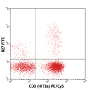
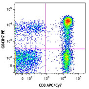
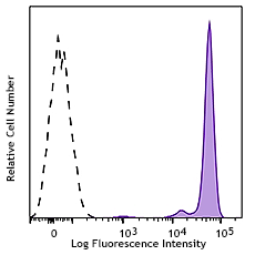
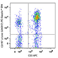
Compare Data Across All Formats
This data display is provided for general comparisons between formats.
Your actual data may vary due to variations in samples, target cells, instruments and their settings, staining conditions, and other factors.
If you need assistance with selecting the best format contact our expert technical support team.


Follow Us