 Login / Register


- Clone
- HI30 (See other available formats)
- Regulatory Status
- RUO
- Workshop
- IV N816
- Other Names
- LCA, T200
- Isotype
- Mouse IgG1, κ
- Ave. Rating
- Submit a Review
- Product Citations
- publications

-

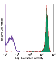
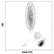
Human peripheral blood lymphocytes stained with HI30 APC
CD45 is a 180-240 kD single chain type I membrane glycoprotein also known as leukocyte common antigen (LCA) and T200. It is a tyrosine phosphatase expressed on the plasma membrane of all hematopoietic cells, except erythrocytes and platelets. CD45 is a signaling molecule that regulates a variety of cellular processes including cell growth, differentiation, cell cycle, and oncogenic transformation. CD45 plays a critical role in T and B cell antigen receptor-mediated activation by dephosphorylating substrates including p56Lck, p59Fyn, and other Src family kinases. CD45 non-covalently associates with lymphocyte phosphatase-associated phosphoprotein (LPAP) on T and B lymphocytes. CD45 has been reported to bind galectin-1 and to be associated with several other cell surface antigens including CD1, CD2, CD3, and CD4.
Product DetailsProduct Details
- Reactivity
- Human
- Antibody Type
- Monoclonal
- Host Species
- Mouse
- Formulation
- Phosphate-buffered solution, pH 7.2, containing 0.09% sodium azide and BSA (origin USA)
- Preparation
- The antibody was purified by affinity chromatography, and conjugated with APC under optimal conditions.
- Concentration
- Lot-specific (to obtain lot-specific concentration and expiration, please enter the lot number in our Certificate of Analysis online tool.)
- Storage & Handling
- The CD45 antibody solution should be stored undiluted between 2°C and 8°C, and protected from prolonged exposure to light. Do not freeze.
- Application
-
FC - Quality tested
- Recommended Usage
-
Each lot of this antibody is quality control tested by immunofluorescent staining with flow cytometric analysis. For flow cytometric staining, the suggested use of this reagent is 5 µl per million cells in 100 µl staining volume or 5 µl per 100 µl of whole blood.
- Excitation Laser
-
Red Laser (633 nm)
- Application Notes
-
Additional reported applications (for the relevant formats) include: immunohistochemical staining of acetone-fixed frozen tissue sections and formalin-fixed paraffin-embedded tissue sections9, inhibition of CD45 functions4, immunofluorescence11, Western blotting3, and spatial biology (IBEX)16,17.
It was found that the HI30 clone and the 2D1 clone can cross block each other's binding. -
Application References
(PubMed link indicates BioLegend citation) -
- Knapp W, et al. 1989. Leucocyte Typing IV. Oxford University Press. New York.
- Kishihara K, et al. 1993. Cell 74:143.
- Esser M, et al. 2001. J. Virol. 75:6173. (WB)
- Yamada T, et al. 2002. J. Biol. Chem. 277:28830.
- Nagano M, et al. 2007. Blood 110:151.
- Jiang Q, et al. 2008. Blood 112:2858. PubMed
- Morozov A, et al. 2010. Clin Cancer Res. 16:5630. PubMed
- Yoshino N, et al. 2000. Exp. Anim. (Tokyo) 49:97. (FC)
- Friedman T, et al. 1999. J. Immunol. 162:5256. (IHC)
- Oeztuerk-Winder F, et al. 2012. EMBO J. 31:3431. (FC) PubMed
- Rees LE, et al. 2003. Clin. Exp. Immunol. 134:497. (IF)
- Lee J, et al. 2015. J Exp Med. 212:385. PubMed
- Breton G, et al. 2015. J Exp Med. 212:401. PubMed
- Marquardt N, et al. 2015. J Immunol. 6:2467. PubMed
- Bushway ME, et al. 2014. Biol Reprod. 90(5): 110. (IF) PubMed
- Radtke AJ, et al. 2020. Proc Natl Acad Sci USA. 117:33455-33465. (SB) PubMed
- Radtke AJ, et al. 2022. Nat Protoc. 17:378-401. (SB) PubMed
- Product Citations
- RRID
-
AB_314399 (BioLegend Cat. No. 304011)
AB_314400 (BioLegend Cat. No. 304012)
AB_2562049 (BioLegend Cat. No. 304037)
Antigen Details
- Structure
- Tyrosine phosphatases, type I transmembrane protein, 180-240 kD (multiple isoforms)
- Distribution
-
Hematopoietic cells, not expressed in circulating erythrocytes or platelets
- Function
- TCR and BCR mediated activation
- Ligand/Receptor
- Galectin-1, CD2, CD3, CD4
- Cell Type
- Hematopoietic stem and progenitors, Mesenchymal Stem Cells
- Biology Area
- Cell Biology, Immunology, Inhibitory Molecules, Innate Immunity, Neuroscience, Neuroscience Cell Markers, Stem Cells
- Molecular Family
- CD Molecules
- Antigen References
-
- Thomas M. 1989. Annu. Rev. Immunol. 7:339.
- Trowbridge I, et al. 1994. Annu. Rev. Immunol.12:85.
- Gene ID
- 5788 View all products for this Gene ID
- UniProt
- View information about CD45 on UniProt.org
Related Pages & Pathways
Pathways


Customers Also Purchased
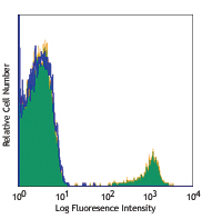
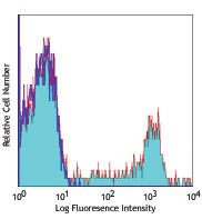
Compare Data Across All Formats
This data display is provided for general comparisons between formats.
Your actual data may vary due to variations in samples, target cells, instruments and their settings, staining conditions, and other factors.
If you need assistance with selecting the best format contact our expert technical support team.


Follow Us