 Login / Register


- Clone
- 9C4 (See other available formats)
- Regulatory Status
- RUO
- Other Names
- Ep-CAM, tumor associated calcium signal transducer 1, epithelial cell surface antigen, epithelial glycoprotein 2, EGP2, adenocarcinoma associated antigen, TROP1
- Isotype
- Mouse IgG2b, κ
- Ave. Rating
- Submit a Review
- Product Citations
- publications

-

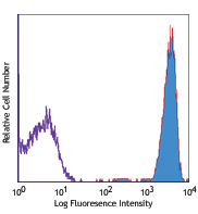
Human colon carcinoma cell line HT29 was stained with 9C4 Brilliant Violet 421™ (filled histogram) or mouse IgG2b, κ Brilliant Violet 421™ isotype control (open histogram).
| Cat # | Size | Price | Quantity Check Availability | Save | ||
|---|---|---|---|---|---|---|
| 324219 | 25 tests | 168€ | ||||
| 324220 | 100 tests | 344€ | ||||
CD326 is also known as Ep-CAM, tumor associated calcium signal transducer 1, epithelial cell surface antigen, epithelial glycoprotein 2, EGP2, adenocarcinoma associated antigen, and TROP1. CD326 is a type I transmembrane protein containing six disulfide bridges and one THYRO domain. This cell surface glycosylated 40 kD protein is highly expressed in bone marrow, colon, lung, and most normal epithelial cells and is expressed on carcinomas of gastrointestinal origin. Recently, it has been reported that CD326 expression occurs during the early steps of erythrogenesis. CD326 functions as a homotypic calcium-independent cell adhesion molecule and is believed to be involved in carcinogenesis by its ability to induce genes involved in cellular metabolism and proliferation. CD326 antigen is an immunotherapeutic target for the treatment of human carcinomas.
Product DetailsProduct Details
- Reactivity
- Human
- Antibody Type
- Monoclonal
- Host Species
- Mouse
- Immunogen
- DU.4475 breast carcinoma
- Formulation
- Phosphate-buffered solution, pH 7.2, containing 0.09% sodium azide and BSA (origin USA).
- Preparation
- The antibody was purified by affinity chromatography and conjugated with Brilliant Violet 421™ under optimal conditions.
- Concentration
- Lot-specific (to obtain lot-specific concentration and expiration, please enter the lot number in our Certificate of Analysis online tool.)
- Storage & Handling
- The antibody solution should be stored undiluted between 2°C and 8°C, and protected from prolonged exposure to light. Do not freeze.
- Application
-
FC - Quality tested
- Recommended Usage
-
Each lot of this antibody is quality control tested by immunofluorescent staining with flow cytometric analysis. For flow cytometric staining, the suggested use of this reagent is 5 µl per million cells in 100 µl staining volume or 5 µl per 100 µl of whole blood.
Brilliant Violet 421™ excites at 405 nm and emits at 421 nm. The standard bandpass filter 450/50 nm is recommended for detection. Brilliant Violet 421™ is a trademark of Sirigen Group Ltd.
Learn more about Brilliant Violet™.
This product is subject to proprietary rights of Sirigen Inc. and is made and sold under license from Sirigen Inc. The purchase of this product conveys to the buyer a non-transferable right to use the purchased product for research purposes only. This product may not be resold or incorporated in any manner into another product for resale. Any use for therapeutics or diagnostics is strictly prohibited. This product is covered by U.S. Patent(s), pending patent applications and foreign equivalents. - Excitation Laser
-
Violet Laser (405 nm)
- Application Notes
-
Additional reported applications (for the revelant formats) include: immunofluorescence, immunohistochemistry3, and spatial biology (IBEX)4,5.
-
Application References
(PubMed link indicates BioLegend citation) -
- Lammers R, et al. 2002. Exp. Hematol. 30:537.
- Schultz LD, et al. 2010. P. Natl. Acad. Sci. USA 107:13022. PubMed
- Human Protein Atlas http://www.proteinatlas.org/ENSG00000119888/antibody (IHC)
- Radtke AJ, et al. 2020. Proc Natl Acad Sci USA. 117:33455-33465. (SB) PubMed
- Radtke AJ, et al. 2022. Nat Protoc. 17:378-401. (SB) PubMed
- Product Citations
- RRID
-
AB_11124342 (BioLegend Cat. No. 324219)
AB_2563847 (BioLegend Cat. No. 324220)
Antigen Details
- Structure
- Type I transmembrane protein, contains six disulfide bridges, one THYRO domain, approximate molecular weight 40 kD.
- Distribution
-
Highly expressed in bone marrow, colon, lung, and most normal epithelial cells. Also highly expressed on carcinomas of gastrointestinal origin. Expressed during early erythrogenesis.
- Function
- Homotypic calcium-independent cell adhesion. CD326 is believed to be involved in carcinogenesis by its ability to induce genes involved in cellular metabolism and proliferation.
- Modification
- Glycosylated.
- Cell Type
- Embryonic Stem Cells, Epithelial cells
- Biology Area
- Cell Biology, Immunology, Stem Cells
- Molecular Family
- Adhesion Molecules, CD Molecules
- Antigen References
-
1. Strnad J, et al. 1989. Cancer Res. 49:314.
2. Munz M, et al. 2004. Oncogene 23:5748.
3. Rao CG, et al. 2005. Int. J. Oncol. 27:49. - Gene ID
- 4072 View all products for this Gene ID
- UniProt
- View information about CD326 on UniProt.org


Related FAQs
- What is the F/P ratio range of our BV421™ format antibody reagents?
-
It is lot-specific. On average it ranges between 2-4.
Customers Also Purchased
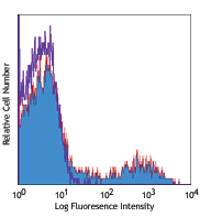
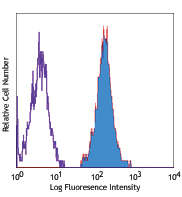
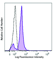
Compare Data Across All Formats
This data display is provided for general comparisons between formats.
Your actual data may vary due to variations in samples, target cells, instruments and their settings, staining conditions, and other factors.
If you need assistance with selecting the best format contact our expert technical support team.


Follow Us