 Login / Register


- Clone
- F38-2E2 (See other available formats)
- Regulatory Status
- RUO
- Workshop
- HCDM listed
- Other Names
- T cell immunoglobulin and mucin domain containing protein 3, hepatitis virus cellular receptor 2, CD366
- Isotype
- Mouse IgG1, κ
- Ave. Rating
- Submit a Review
- Product Citations
- publications

-

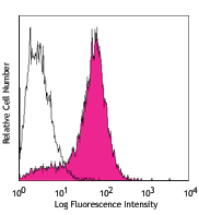
Th1-polarized human peripheral blood lymphocytes were stained with anti-human CD366 (Tim-3, clone F38-2E2) Brilliant Violet 421™ (filled histogram) or mouse IgG1, κ Brilliant Violet 421™ isotype control (open histogram).
| Cat # | Size | Price | Quantity Check Availability | Save | ||
|---|---|---|---|---|---|---|
| 345007 | 25 tests | 172€ | ||||
| 345008 | 100 tests | 326€ | ||||
CD366 (Tim-3) is a transmembrane protein also known as T cell immunoglobulin and mucin domain containing protein-3. Tim-3 is expressed at high levels on activated T cells (preferentially on Th1 cells, monocytes/macrophages, and dendritic cells). Tim-3 has also been shown to exist as a soluble protein. Cells expressing Tim-3 are present at high levels in the CNS of animals at the onset of experimental autoimmune encephalomyelitis (EAE), a disease mediated by lymphocytes secreting Th1-like cytokines. Tim-3 has been proposed to inhibit Th1-mediated immune responses and promote immunological tolerance.
Product DetailsProduct Details
- Reactivity
- Human
- Antibody Type
- Monoclonal
- Host Species
- Mouse
- Immunogen
- Human Tim-3 fusion protein
- Formulation
- Phosphate-buffered solution, pH 7.2, containing 0.09% sodium azide and BSA (origin USA).
- Preparation
- The antibody was purified by affinity chromatography and conjugated with Brilliant Violet 421™ under optimal conditions.
- Concentration
- Lot-specific (to obtain lot-specific concentration and expiration, please enter the lot number in our Certificate of Analysis online tool.)
- Storage & Handling
- The antibody solution should be stored undiluted between 2°C and 8°C, and protected from prolonged exposure to light. Do not freeze.
- Application
-
FC - Quality tested
- Recommended Usage
-
Each lot of this antibody is quality control tested by immunofluorescent staining with flow cytometric analysis. For flow cytometric staining, the suggested use of this reagent is 5 µl per million cells in 100 µl staining volume or 5 µl per 100 µl of whole blood.
Brilliant Violet 421™ excites at 405 nm and emits at 421 nm. The standard bandpass filter 450/50 nm is recommended for detection. Brilliant Violet 421™ is a trademark of Sirigen Group Ltd.
Learn more about Brilliant Violet™.
This product is subject to proprietary rights of Sirigen Inc. and is made and sold under license from Sirigen Inc. The purchase of this product conveys to the buyer a non-transferable right to use the purchased product for research purposes only. This product may not be resold or incorporated in any manner into another product for resale. Any use for therapeutics or diagnostics is strictly prohibited. This product is covered by U.S. Patent(s), pending patent applications and foreign equivalents. - Excitation Laser
-
Violet Laser (405 nm)
- Application Notes
-
Additional reported applications (for relevant formats of this clone) include: costimulation1 (clone 2E2 has been shown to enhance T-cell receptor mediated activation and cytokine secretion) and blocking2,3.
-
Application References
(PubMed link indicates BioLegend citation) -
- Hastings WD, et al. 2009. Eur. J. Immunol. 39:2492. (Costim)
- Jones RB, et al. 2008. J. Exp. Med. 205:2763. (Block)
- Klibi J, et al 2009. Blood 113:1957. (FC, Block)
- Product Citations
- RRID
-
AB_10900073 (BioLegend Cat. No. 345007)
AB_11218598 (BioLegend Cat. No. 345008)
Antigen Details
- Structure
- Transmembrane protein containing immunoglobulin domain and mucin-like domain; can exist as a soluble form lacking mucin and transmembrane domains
- Distribution
-
Activated T cells, preferentially on Th1 cells, monocytes, dendritic cells
- Function
- Plays a role in regulating macrophage activation, T cell apoptosis and immune tolerance
- Ligand/Receptor
- Galectin-9
- Cell Type
- Dendritic cells, Monocytes, T cells, Th1, Tregs
- Biology Area
- Immunology, Inhibitory Molecules
- Molecular Family
- CD Molecules, Immune Checkpoint Receptors
- Antigen References
-
1. Hafler DA and Kuchroo V. 2008. J. Exp. Med. 205:2699.
2. Zhu C, et al. 2005. Nat. Immunol. 6:1245.
3. Wang F, et al. 2009. Immunobiology 214:342. - Gene ID
- 84868 View all products for this Gene ID
- UniProt
- View information about CD366 on UniProt.org


Related FAQs
- What is the F/P ratio range of our BV421™ format antibody reagents?
-
It is lot-specific. On average it ranges between 2-4.
Customers Also Purchased
Compare Data Across All Formats
This data display is provided for general comparisons between formats.
Your actual data may vary due to variations in samples, target cells, instruments and their settings, staining conditions, and other factors.
If you need assistance with selecting the best format contact our expert technical support team.


Follow Us