 Login / Register


- Clone
- A15153G (See other available formats)
- Regulatory Status
- RUO
- Other Names
- T-cell immunoreceptor with Ig and ITIM domains, VSIG9, VSTM3, WUCAM
- Isotype
- Mouse IgG2a, κ
- Ave. Rating
- Submit a Review
- Product Citations
- publications

-

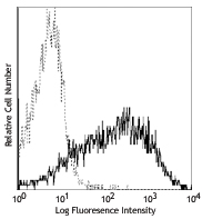
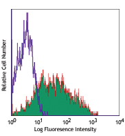
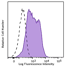
Human peripheral blood leukocytes were stained with CD3 FITC and TIGIT (clone A15153G) Brilliant Violet 605™ (top) or mouse IgG2a, κ Brilliant Violet 605™ isotype control (bottom). Data shown was gated on the lymphocyte population. -

| Cat # | Size | Price | Quantity Check Availability | Save | ||
|---|---|---|---|---|---|---|
| 372711 | 25 tests | 172€ | ||||
| 372712 | 100 tests | 357€ | ||||
T cell immunoreceptor with Ig and ITIM domains (TIGIT), also known as VSTM3 or WUCAM, is a 26 kD, type I transmembrane protein and is a member of the PVR (poliovirus receptor) family of immunoglobulin-like domain containing proteins. TIGIT is expressed on activated T cells, follicular T helper, memory, and regulatory T cells as well as on NK cells. TIGIT is a negative regulator of NK and T cell activation. Expression of TIGIT is associated with decreased functionality of CD8 T cells in chronic viral infection and tumors. TIGIT also promotes the differentiation of tolerogenic phenotype in dendritic cells with an increased secretion of IL-10 and a diminished production of IL-12.
Product DetailsProduct Details
- Reactivity
- Human
- Antibody Type
- Monoclonal
- Host Species
- Mouse
- Immunogen
- Recombinant Human TIGIT.
- Formulation
- Phosphate-buffered solution, pH 7.2, containing 0.09% sodium azide and BSA (origin USA).
- Preparation
- The antibody was purified by affinity chromatography and conjugated with Brilliant Violet 605™ under optimal conditions.
- Concentration
- Lot-specific (to obtain lot-specific concentration and expiration, please enter the lot number in our Certificate of Analysis online tool.)
- Storage & Handling
- The antibody solution should be stored undiluted between 2°C and 8°C, and protected from prolonged exposure to light. Do not freeze.
- Application
-
FC - Quality tested
- Recommended Usage
-
Each lot of this antibody is quality control tested by immunofluorescent staining with flow cytometric analysis. For flow cytometric staining, the suggested use of this reagent is 5 µl per million cells in 100 µl staining volume or 5 µl per 100 µl of whole blood.
Brilliant Violet 605™ excites at 405 nm and emits at 603 nm. The bandpass filter 610/20 nm is recommended for detection, although filter optimization may be required depending on other fluorophores used. Be sure to verify that your cytometer configuration and software setup are appropriate for detecting this channel. Refer to your instrument manual or manufacturer for support. Brilliant Violet 605™ is a trademark of Sirigen Group Ltd.
Learn more about Brilliant Violet™.
This product is subject to proprietary rights of Sirigen Inc. and is made and sold under license from Sirigen Inc. The purchase of this product conveys to the buyer a non-transferable right to use the purchased product for research purposes only. This product may not be resold or incorporated in any manner into another product for resale. Any use for therapeutics or diagnostics is strictly prohibited. This product is covered by U.S. Patent(s), pending patent applications and foreign equivalents. - Excitation Laser
-
Violet Laser (405 nm)
- Application Notes
-
This clone can suppress anti-CD3 induced T cell proliferation in vitro based on in-house testing.
This clone has been tested in-house and determined to not be suitable for applications in immunohistochemistry of paraffin-embedded tissue sections (IHC-P).
Additional reported applications (for the relevant formats) include: Blocking1. -
Application References
(PubMed link indicates BioLegend citation) -
- Stamm H, et al. 2018. Oncogene. Pubmed
- Product Citations
- RRID
-
AB_2632926 (BioLegend Cat. No. 372711)
AB_2632927 (BioLegend Cat. No. 372712)
Antigen Details
- Structure
- 26kD; type I transmembrane protein, Ig-like V-type domain, ITIM motif.
- Distribution
-
Activated T cells, Regulatory T cells (Treg), Follicular Helper T cells (TFH), NK cells.
- Function
- Cell signaling, negative regulation of T cells, T cell tolerance, T cell anergy.
- Ligand/Receptor
- CD155 (PVR), CD112 (PVRL2, NECTIN-2).
- Cell Type
- NK cells, T cells, Tfh, Tregs
- Biology Area
- Cell Adhesion, Cell Biology, Immunology, Inhibitory Molecules, Signal Transduction
- Molecular Family
- Adhesion Molecules, Immune Checkpoint Receptors
- Antigen References
-
1. Stanietsky N, et al. 2009. Proc. Natl. Acad. Sci. 106:17858.
2. Yu X, et al. 2009. Nat. Immunol. 10:48.
3. Johnston R, et al. 2014. Cancer Cell. 26:923. - Gene ID
- 201633 View all products for this Gene ID
- UniProt
- View information about TIGIT on UniProt.org


Related FAQs
Customers Also Purchased
Compare Data Across All Formats
This data display is provided for general comparisons between formats.
Your actual data may vary due to variations in samples, target cells, instruments and their settings, staining conditions, and other factors.
If you need assistance with selecting the best format contact our expert technical support team.


Follow Us