 Login / Register


- Clone
- HIT2 (See other available formats)
- Regulatory Status
- RUO
- Workshop
- III 155
- Other Names
- T10, ADP-ribosyl cyclase
- Isotype
- Mouse IgG1, κ
- Ave. Rating
- Submit a Review
- Product Citations
- publications

-

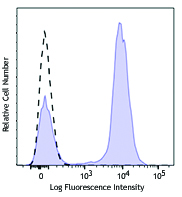
Human peripheral blood lymphocytes were stained with CD38 (clone HIT2) Brilliant Violet 711™ (filled histogram) or mouse IgG1, κ Brilliant Violet 711™ isotype control (open histogram).
| Cat # | Size | Price | Quantity Check Availability | Save | ||
|---|---|---|---|---|---|---|
| 303527 | 25 tests | 185€ | ||||
| 303528 | 100 tests | 317€ | ||||
CD38 is a 45 kD type II transmembrane glycoprotein also known as T10. It is an ADP-ribosyl hydrolase expressed at variable levels on hematopoietic cells and in some non-hematopoietic tissues (such as brain, muscles, and kidney). In humans, it is expressed at high levels on plasma cells and activated T and B cells. By functioning as both a cyclase and a hydrolase, CD38 mediates lymphocyte activation, adhesion, and the metabolism of cADPR and NAADP. CD31 is the ligand of CD38.
Product DetailsProduct Details
- Reactivity
- Human
- Antibody Type
- Monoclonal
- Host Species
- Mouse
- Formulation
- Phosphate-buffered solution, pH 7.2, containing 0.09% sodium azide and BSA (origin USA).
- Preparation
- The antibody was purified by affinity chromatography and conjugated with Brilliant Violet 711™ under optimal conditions.
- Concentration
- Lot-specific (to obtain lot-specific concentration and expiration, please enter the lot number in our Certificate of Analysis online tool.)
- Storage & Handling
- The antibody solution should be stored undiluted between 2°C and 8°C, and protected from prolonged exposure to light. Do not freeze.
- Application
-
FC - Quality tested
- Recommended Usage
-
Each lot of this antibody is quality control tested by immunofluorescent staining with flow cytometric analysis. For flow cytometric staining, the suggested use of this reagent is 5 µl per million cells in 100 µl staining volume or 5 µl per 100 µl of whole blood.
Brilliant Violet 711™ excites at 405 nm and emits at 711 nm. The bandpass filter 710/50 nm is recommended for detection, although filter optimization may be required depending on other fluorophores used. Be sure to verify that your cytometer configuration and software setup are appropriate for detecting this channel. Refer to your instrument manual or manufacturer for support. Brilliant Violet 711™ is a trademark of Sirigen Group Ltd.
Learn more about Brilliant Violet™.
This product is subject to proprietary rights of Sirigen Inc. and is made and sold under license from Sirigen Inc. The purchase of this product conveys to the buyer a non-transferable right to use the purchased product for research purposes only. This product may not be resold or incorporated in any manner into another product for resale. Any use for therapeutics or diagnostics is strictly prohibited. This product is covered by U.S. Patent(s), pending patent applications and foreign equivalents. - Excitation Laser
-
Violet Laser (405 nm)
- Application Notes
-
Additional reported applications (for the relevant formats) include: immunohistochemical staining of acetone-fixed frozen tissue sections6 and spatial biology (IBEX)10,11.
-
Application References
(PubMed link indicates BioLegend citation) -
- Kishimoto T, et al. Eds. 1997. Leucocyte Typing VI. Garland Publishing Inc. London.
- Dieu M. 1998. J. Exp. Med. 188:373.
- Esser M, et al. 2001. J. Virol. 75:6173.
- Jeannin P, et al. 1999. J. Immunol. 162:2044.
- Kapsogeorgou EK, et al. 2001. J. Immunol. 166:3107.
- van der Voort R, et al. 1997. J. Exp. Med. 185:2121. (IHC)
- Bende RJ, et al. 2003. Am. J. Pathol. 162:105.
- Lehner M, et al. 2008. J. Leukoc. Biol. 83:883. PubMed
- Yoshino N, et al. 2000. Exp. Anim. (Tokyo) 49:97. (FC)
- Radtke AJ, et al. 2020. Proc Natl Acad Sci USA. 117:33455-33465. (SB) PubMed
- Radtke AJ, et al. 2022. Nat Protoc. 17:378-401. (SB) PubMed
- Product Citations
- RRID
-
AB_11218990 (BioLegend Cat. No. 303527)
AB_2563811 (BioLegend Cat. No. 303528)
Antigen Details
- Structure
- ADP-ribosyl cyclase, ectoenzyme, type II glycoprotein, 45 kD
- Distribution
-
T cells, B cells, NK, myeloid, plasma, and dendritic cells
- Function
- Ecto-ADP-ribosyl cyclase, calcium signaling, cell activation
- Ligand/Receptor
- CD31, hyaluronic acid
- Cell Type
- B cells, Dendritic cells, NK cells, Plasma cells, T cells
- Biology Area
- Immunology
- Molecular Family
- Adhesion Molecules, CD Molecules
- Antigen References
-
1. Ferrero E, et al. 1999. J. Leukoc. Biol. 65:151.
2. Lund F, et al. 1995. Immunol. Today 16:469. - Gene ID
- 952 View all products for this Gene ID
- UniProt
- View information about CD38 on UniProt.org
Related Pages & Pathways
Pathways


Customers Also Purchased
Compare Data Across All Formats
This data display is provided for general comparisons between formats.
Your actual data may vary due to variations in samples, target cells, instruments and their settings, staining conditions, and other factors.
If you need assistance with selecting the best format contact our expert technical support team.


Follow Us