 Login / Register


- Clone
- N418 (See other available formats)
- Regulatory Status
- RUO
- Other Names
- αX integrin, integrin αX chain, CR4, p150, ITGAX
- Isotype
- Armenian Hamster IgG
- Ave. Rating
- Submit a Review
- Product Citations
- publications

-

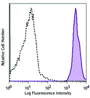
C57BL/6 mouse splenocytes were stained with I-A/I-E FITC and CD11c (clone N418) Brilliant Violet 785™.
| Cat # | Size | Price | Quantity Check Availability | Save | ||
|---|---|---|---|---|---|---|
| 117335 | 125 µL | 172€ | ||||
| 117336 | 50 µg | 229€ | ||||
CD11c is a 150 kD glycoprotein also known as αX integrin, CR4, and p150. CD11c forms a αXβ2 heterodimer with β2 integrin (CD18). It is primarily expressed on dendritic cells, NK cells, a subset of intestinal intraepithelial lymphocytes (IEL), and some activated T cells. The αXβ2 integrin plays an important role in cell-cell contact by binding its ligands: iC3b, fibrinogen, and CD54.
Product DetailsProduct Details
- Reactivity
- Mouse
- Antibody Type
- Monoclonal
- Host Species
- Armenian Hamster
- Immunogen
- Mouse spleen dendritic cells
- Formulation
- Phosphate-buffered solution, pH 7.2, containing 0.09% sodium azide and BSA (origin USA).
- Preparation
- The antibody was purified by affinity chromatography and conjugated with Brilliant Violet 785™ under optimal conditions.
- Concentration
- µg sizes: 0.2 mg/mLµL sizes: lot-specific (to obtain lot-specific concentration and expiration, please enter the lot number in our Certificate of Analysis online tool.)
- Storage & Handling
- The antibody solution should be stored undiluted between 2°C and 8°C, and protected from prolonged exposure to light. Do not freeze.
- Application
-
FC - Quality tested
- Recommended Usage
-
Each lot of this antibody is quality control tested by immunofluorescent staining with flow cytometric analysis. For flow cytometric staining using the µg size, the suggested use of this reagent is ≤0.25 µg per million cells in 100 µl volume. For flow cytometric staining using the µl size, the suggested use of this reagent is 5 µl per million cells in 100 µl staining volume or 5 µl per 100 µl of whole blood. It is recommended that the reagent be titrated for optimal performance for each application.
Brilliant Violet 785™ excites at 405 nm and emits at 785 nm. The bandpass filter 780/60 nm is recommended for detection, although filter optimization may be required depending on other fluorophores used. Be sure to verify that your cytometer configuration and software setup are appropriate for detecting this channel. Refer to your instrument manual or manufacturer for support. Brilliant Violet 785™ is a trademark of Sirigen Group Ltd.
Learn more about Brilliant Violet™.
This product is subject to proprietary rights of Sirigen Inc. and is made and sold under license from Sirigen Inc. The purchase of this product conveys to the buyer a non-transferable right to use the purchased product for research purposes only. This product may not be resold or incorporated in any manner into another product for resale. Any use for therapeutics or diagnostics is strictly prohibited. This product is covered by U.S. Patent(s), pending patent applications and foreign equivalents. - Excitation Laser
-
Violet Laser (405 nm)
- Application Notes
-
Additional reported applications (for the relevant formats) include: immunoprecipitation3, immunohistochemical staining of acetone-fixed frozen sections3, immunofluorescence microscopy5, 9 (Alexa Fluor® 488 conjugated N418 was used for IHC in frozen sections10), and spatial biology (IBEX)22,23.
-
Application References
(PubMed link indicates BioLegend citation) -
- Granucci F, et al. 1997. J. Immunol. 159:1794.
- Stokes RW, et al. 1998. J. Immunol. 160:5514.
- Metlay JP, et al. 1990. J. Exp. Med. 171:1753. (IHC, IP)
- Ma XT, et al. 2006. Cancer Research 66:1169.
- Chin RK, et al. 2006. J. Immunol. 177:290. (IF)
- Cervantes-Barragan L, et al. 2007. Blood 109:1131. (FC) PubMed
- Turnquist HR, et al. 2007. J. Immunol. 178:7018. (FC) PubMed
- Benson MJ, et al. 2007. J. Exp. Med. doi:10.1084/jem.20070719. (FC) PubMed
- You Y, et al. 2009. J. Immunol. 182:7343. (IF) PubMed
- Roland CL, et al. 2009. Mol. Cancer Res. 8:1761. (IHC, FC) PubMed
- Wikstrom M, et al.2006. J. Immunol. 177:913. PubMed
- Pericolini E, et al. 2008. J. Leukocyte Biol. 83:1286. PubMed
- Randall LM, et al. 2008. Infect. Immun.76:3312. PubMed
- Fahlen-Yrild L, et al. 2009. J. Immunol. 183:5032. PubMed
- Osterholzer JJ, et al. 2009. J. Immunol. 183:8044. PubMed
- Bankoti J, et al. 2010. Toxicol. Sci. 115:422. (FC) PubMed
- Eisenach PA, et al. 2010. J Cell Sci. 123:4182. PubMed
- Leppin K, et al. 2014. Invest. Ophthalmol. Vis. Sci. 55:3603. PubMed
- Sakai F, et al. 2014. PLoS One. 9:105370. PubMed
- Gibbins JD, et al. 2014. Blood. 124:2953. PubMed
- White CE, et al. 2015. J Immunol. 194:697. PubMed
- Lu X, et al. 2015. J Immunol. 194:2011. PubMed
- Radtke AJ, et al. 2020. Proc Natl Acad Sci U S A. 117:33455-65. (SB) PubMed
- Radtke AJ, et al. 2022. Nat Protoc. 17:378-401. (SB) PubMed
- Product Citations
- RRID
-
AB_11219204 (BioLegend Cat. No. 117335)
AB_2565268 (BioLegend Cat. No. 117336)
Antigen Details
- Structure
- Integrin α-chain, associates with integrin β2 (CD18), 150 kD
- Distribution
-
Dendritic cells, NK cells, intestinal intraepithelial lymphocytes (IEL), some activated T cells
- Function
- Cellular adhesion
- Ligand/Receptor
- iC3b, fibrinogen
- Cell Type
- Dendritic cells, Epithelial cells, NK cells, T cells, Tregs
- Biology Area
- Cell Adhesion, Cell Biology, Costimulatory Molecules, Immunology, Innate Immunity, Neuroscience, Neuroscience Cell Markers
- Molecular Family
- Adhesion Molecules, CD Molecules
- Antigen References
-
1. Barclay A, et al. 1997. The Leukocyte Antigen Facts Book Academic Press.
2. Springer TA. 1994. Cell 76:301.
3. Lopez-Rodriguez C, et al. 1996. J. Immunol. 156:3780. - Gene ID
- 16411 View all products for this Gene ID
- UniProt
- View information about CD11c on UniProt.org
Related Pages & Pathways
Pathways


Customers Also Purchased
Compare Data Across All Formats
This data display is provided for general comparisons between formats.
Your actual data may vary due to variations in samples, target cells, instruments and their settings, staining conditions, and other factors.
If you need assistance with selecting the best format contact our expert technical support team.


Follow Us