 Login / Register


- Clone
- 30-H12 (See other available formats)
- Regulatory Status
- RUO
- Other Names
- Thy-1.2
- Isotype
- Rat IgG2b, κ
- Ave. Rating
- Submit a Review
- Product Citations
- publications

-

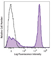
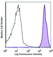
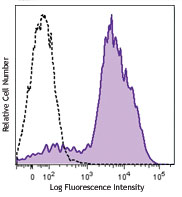
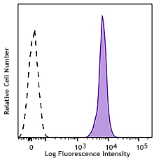
C57BL/6 thymocytes were stained with CD90.2 (clone 30-H12) Brilliant Violet 785™ (filled histogram) or rat IgG2b, κ Brilliant Violet 785™ isotype control (open histogram).
| Cat # | Size | Price | Quantity Check Availability | Save | ||
|---|---|---|---|---|---|---|
| 105331 | 50 µg | 248€ | ||||
CD90.2 is a 25-35 kD immunoglobulin superfamily member also known as Thy1.2. It is expressed on hematopoietic stem cells and neurons, all thymocytes, and peripheral T cells in Thy1.2 bearing mouse strains (Balb/c, CBA/J, C3H/He, C57BL/-, DBA, NZB/-). CD90.2 is a glycosylphosphatidylinositol (GPI)-anchored membrane glycoprotein involved in signal transduction. CD90.2 is involved in costimulation of lymphocyte proliferation and induction of hematopoietic stem cells differentiation. CD90.2 has been shown to interact with CD45. The 30-H12 antibody has been reported to induce Ca2+ flux in thymocytes and, in combination with antibody against the CD3/TCR complex, promote thymocyte apoptosis and inhibit CD3-mediated proliferative responses of mature T lymphocytes.
Product DetailsProduct Details
- Reactivity
- Mouse
- Antibody Type
- Monoclonal
- Host Species
- Rat
- Immunogen
- Mouse thymus or spleen
- Formulation
- Phosphate-buffered solution, pH 7.2, containing 0.09% sodium azide and BSA (origin USA).
- Preparation
- The antibody was purified by affinity chromatography and conjugated with Brilliant Violet 785™ under optimal conditions.
- Concentration
- 0.2 mg/ml
- Storage & Handling
- The antibody solution should be stored undiluted between 2°C and 8°C, and protected from prolonged exposure to light. Do not freeze.
- Application
-
FC - Quality tested
- Recommended Usage
-
Each lot of this antibody is quality control tested by immunofluorescent staining with flow cytometric analysis. For flow cytometric staining, the suggested use of this reagent is ≤0.5 µg per million cells in 100 µl volume. It is recommended that the reagent be titrated for optimal performance for each application.
Brilliant Violet 785™ excites at 405 nm and emits at 785 nm. The bandpass filter 780/60 nm is recommended for detection, although filter optimization may be required depending on other fluorophores used. Be sure to verify that your cytometer configuration and software setup are appropriate for detecting this channel. Refer to your instrument manual or manufacturer for support. Brilliant Violet 785™ is a trademark of Sirigen Group Ltd.
Learn more about Brilliant Violet™.
This product is subject to proprietary rights of Sirigen Inc. and is made and sold under license from Sirigen Inc. The purchase of this product conveys to the buyer a non-transferable right to use the purchased product for research purposes only. This product may not be resold or incorporated in any manner into another product for resale. Any use for therapeutics or diagnostics is strictly prohibited. This product is covered by U.S. Patent(s), pending patent applications and foreign equivalents. - Excitation Laser
-
Violet Laser (405 nm)
- Application Notes
-
Additional reported applications (for the relevant formats) include: in vivo and in vitro depletion1,2,7, costimulation of CD3/TCR-mediated signal transduction3,4, and immunohistochemical staining5 of acetone-fixed frozen sections. The 30-H12 antibody does not react with Thy-1.1 alloantigen of the AKR/J and PL strains. To reduce non-specific binding to cells bearing Fc-receptors, pre-incubation of cells with anti-mouse CD16/CD32, clone 93 (Cat. No. 101301 & 101302) is recommended prior to immunofluorescent staining. The Ultra-LEAF™ purified antibody (Endotoxin <0.01 EU/µg, Azide-Free, 0.2 µm filtered) is recommended for functional assays (Cat. Nos. 105351 & 105352).
-
Application References
(PubMed link indicates BioLegend citation) -
- Hathcock KS. 1991. Current Protocols in Immunology. 3.4.1. (Deplete)
- Seaman WE. 1983. J. Immunol. 130:1713. (Deplete)
- Nakashima I, et al. 1991. J. Immunol. 147:1153. (Costim)
- Nakashima I, et al. 1993. J. Immunol. 151:3511. (Costim)
- Ledbetter JA, et al. 1980. J. Exp. Med. 152:280. (IHC)
- Hardy B, et al. 2005. Int. Immunol. 17:615.
- Drobyski W, et al. 1996. Blood 87:5355. (Deplete)
- Dyer KD, et al. 2007. J. Immunol. 179:1693. (FC) PubMed
- Sungur CM, et al. 2013. PNAS. 110:7401. PubMed
- Product Citations
- RRID
-
AB_2562900 (BioLegend Cat. No. 105331)
Antigen Details
- Structure
- Ig superfamily, 25-35 kD
- Distribution
-
Hematopoietic stem cells and neurons, all thymocytes, peripheral T cells of the Thy-1.2 bearing mice
- Function
- Lymphocyte costimulation, proliferation and differentiation of hematopoietic stem cells
- Ligand/Receptor
- CD45
- Cell Type
- Hematopoietic stem and progenitors, Neurons, T cells, Thymocytes
- Biology Area
- Immunology
- Molecular Family
- CD Molecules
- Antigen References
-
1. Barclay A, et al. 1997. The Leukocyte Antigen FactsBook Academic Press.
2. Craig W, et al. 1993. J. Exp. Med. 177:1331.
3. Reif AE and Schlesinger M. 1989. Cell Surface Antigen Thy-1.
4. Mayani H, et al. 1994. Blood 83:2410. - Gene ID
- 21838 View all products for this Gene ID
- UniProt
- View information about CD90.2 on UniProt.org


Customers Also Purchased
Compare Data Across All Formats
This data display is provided for general comparisons between formats.
Your actual data may vary due to variations in samples, target cells, instruments and their settings, staining conditions, and other factors.
If you need assistance with selecting the best format contact our expert technical support team.


Follow Us