 Login / Register


- Clone
- IA6-2 (See other available formats)
- Regulatory Status
- RUO
- Other Names
- Ig delta chain C region
- Isotype
- Mouse IgG2a, κ
- Ave. Rating
- Submit a Review
- Product Citations
- publications


| Cat # | Size | Price | Quantity Check Availability | Save | ||
|---|---|---|---|---|---|---|
| 348205 | 25 tests | 87€ | ||||
| 348206 | 100 tests | 202€ | ||||
IgD, a member of the immunoglobulin (Ig) family, is expressed in naïve B cells. It has 3 Ig-like domains and exists in a transmembrane and a soluble form. In general, IgD is not secreted and usually its expression is lost after the Ig isotype switch. After antigen binding, IgD signals through the CD79a/CD79b (Igα/Igβ) heterodimer, resulting in the activation of the B cell.
Product DetailsProduct Details
- Reactivity
- Human
- Antibody Type
- Monoclonal
- Host Species
- Mouse
- Immunogen
- Human IgD
- Formulation
- Phosphate-buffered solution, pH 7.2, containing 0.09% sodium azide and BSA (origin USA)
- Preparation
- The antibody was purified by affinity chromatography, and conjugated with FITC under optimal conditions.
- Concentration
- Lot-specific (to obtain lot-specific concentration and expiration, please enter the lot number in our Certificate of Analysis online tool.)
- Storage & Handling
- The antibody solution should be stored undiluted between 2°C and 8°C, and protected from prolonged exposure to light. Do not freeze.
- Application
-
FC - Quality tested
- Recommended Usage
-
Each lot of this antibody is quality control tested by immunofluorescent staining with flow cytometric analysis. For flow cytometric staining, the suggested use of this reagent is 5 µl per million cells in 100 µl staining volume or 5 µl per 100 µl of whole blood.
- Excitation Laser
-
Blue Laser (488 nm)
- Application Notes
-
Additional reported applications (for the relevant formats) include: immunohistochemical staining of paraformaldehyde fixed frozen sections4 and spatial biology (IBEX)5,6.
-
Application References
(PubMed link indicates BioLegend citation) -
- Chen K, et al. 2009. Nat. Immunol. 10:889.
- Lee CH, et al. 2005. J. Exp. Med. 203:63.
- Sutter JA, et al. 2008. Clin. Immunol. 126:282.
- Li H and Pauza CD. 2015. Eur. J. Immunol. 45:298. (IHC)
- Radtke AJ, et al. 2020. Proc Natl Acad Sci USA. 117:33455-33465. (SB) PubMed
- Radtke AJ, et al. 2022. Nat Protoc. 17:378-401. (SB) PubMed
- Product Citations
- RRID
-
AB_10613638 (BioLegend Cat. No. 348205)
AB_10612567 (BioLegend Cat. No. 348206)
Antigen Details
- Structure
- Exists in a transmembranal and a soluble form
- Distribution
-
Naïve B cells
- Function
- Antigen binding, B cell activation
- Interaction
- The CD79a/CD79b heterodimer
- Cell Type
- B cells
- Biology Area
- Immunology
- Antigen References
-
1. Geisberger R, et al. 2006. Immunology 118:429.
2. Weller S, et al. 2005. Eur. J. Immunol. 35:2789.
3. Brandtzaeg P and Johansen FE. 2005. Immunol. Rev. 206:32. - Gene ID
- 3495 View all products for this Gene ID
- UniProt
- View information about IgD on UniProt.org
Related Pages & Pathways
Pathways


Related FAQs
Customers Also Purchased
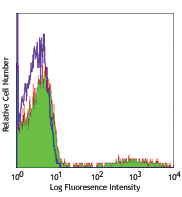
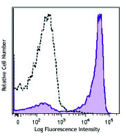
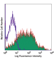
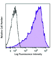
Compare Data Across All Formats
This data display is provided for general comparisons between formats.
Your actual data may vary due to variations in samples, target cells, instruments and their settings, staining conditions, and other factors.
If you need assistance with selecting the best format contact our expert technical support team.


Follow Us