 Login / Register


- Clone
- N418 (See other available formats)
- Regulatory Status
- RUO
- Other Names
- αX integrin, integrin αX chain, CR4, p150, ITGAX
- Isotype
- Armenian Hamster IgG
- Ave. Rating
- Submit a Review
- Product Citations
- publications

-

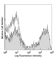
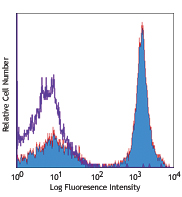
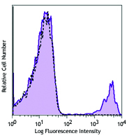
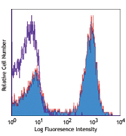
C57BL/6 mouse splenocytes stained with APC anti-mouse I-A/I-E (clone M5/114.15.2) and biotinylated N418 (top) or biotinylated Armenian hamster IgG isotype control (bottom), followed by Sav-PE -

-

Frozen mouse spleen sections. Endogenous biotin blocking applied prior to incubation of primary antibody anti‐mouse CD11c biotin followed by secondary detection with Alexa Fluor® 555 streptavidin. Images were acquired with an automated widefield microscope (Nikon Eclipse Ti) and a CCD camera (QImaging Retiga 2000R). Images provided by Ann Haberman and Tingting Zhang, Yale University.
| Cat # | Size | Price | Quantity Check Availability | Save | ||
|---|---|---|---|---|---|---|
| 117303 | 50 µg | 62€ | ||||
| 117304 | 500 µg | 212€ | ||||
CD11c is a 150 kD glycoprotein also known as αX integrin, CR4, and p150. CD11c forms a αXβ2 heterodimer with β2 integrin (CD18). It is primarily expressed on dendritic cells, NK cells, a subset of intestinal intraepithelial lymphocytes (IEL), and some activated T cells. The αXβ2 integrin plays an important role in cell-cell contact by binding its ligands: iC3b, fibrinogen, and CD54.
Product DetailsProduct Details
- Reactivity
- Mouse
- Antibody Type
- Monoclonal
- Host Species
- Armenian Hamster
- Immunogen
- Mouse spleen dendritic cells
- Formulation
- Phosphate-buffered solution, pH 7.2, containing 0.09% sodium azide.
- Preparation
- The antibody was purified by affinity chromatography, and conjugated with biotin under optimal conditions.
- Concentration
- 0.5 mg/mL
- Storage & Handling
- The antibody solution should be stored undiluted between 2°C and 8°C. Do not freeze.
- Application
-
FC - Quality tested
IHC-F - Verified - Recommended Usage
-
Each lot of this antibody is quality control tested by immunofluorescent staining with flow cytometric analysis. For flow cytometric staining, the suggested use of this reagent is ≤ 0.25 µg per 106 cells in 100 µL volume. It is recommended that the reagent be titrated for optimal performance for other applications.
- Application Notes
-
Additional reported applications (for the relevant formats) include: immunoprecipitation3, immunohistochemical staining of acetone-fixed frozen sections3, immunofluorescence microscopy5, 9 (Alexa Fluor® 488 conjugated N418 was used for IHC in frozen sections10), and spatial biology (IBEX)22,23.
-
Application References
(PubMed link indicates BioLegend citation) -
- Granucci F, et al. 1997. J. Immunol. 159:1794.
- Stokes RW, et al. 1998. J. Immunol. 160:5514.
- Metlay JP, et al. 1990. J. Exp. Med. 171:1753. (IHC, IP)
- Ma XT, et al. 2006. Cancer Research 66:1169.
- Chin RK, et al. 2006. J. Immunol. 177:290. (IF)
- Cervantes-Barragan L, et al. 2007. Blood 109:1131. (FC) PubMed
- Turnquist HR, et al. 2007. J. Immunol. 178:7018. (FC) PubMed
- Benson MJ, et al. 2007. J. Exp. Med. doi:10.1084/jem.20070719. (FC) PubMed
- You Y, et al. 2009. J. Immunol. 182:7343. (IF) PubMed
- Roland CL, et al. 2009. Mol. Cancer Res. 8:1761. (IHC, FC) PubMed
- Wikstrom M, et al.2006. J. Immunol. 177:913. PubMed
- Pericolini E, et al. 2008. J. Leukocyte Biol. 83:1286. PubMed
- Randall LM, et al. 2008. Infect. Immun.76:3312. PubMed
- Fahlen-Yrild L, et al. 2009. J. Immunol. 183:5032. PubMed
- Osterholzer JJ, et al. 2009. J. Immunol. 183:8044. PubMed
- Bankoti J, et al. 2010. Toxicol. Sci. 115:422. (FC) PubMed
- Eisenach PA, et al. 2010. J Cell Sci. 123:4182. PubMed
- Leppin K, et al. 2014. Invest. Ophthalmol. Vis. Sci. 55:3603. PubMed
- Sakai F, et al. 2014. PLoS One. 9:105370. PubMed
- Gibbins JD, et al. 2014. Blood. 124:2953. PubMed
- White CE, et al. 2015. J Immunol. 194:697. PubMed
- Lu X, et al. 2015. J Immunol. 194:2011. PubMed
- Radtke AJ, et al. 2020. Proc Natl Acad Sci U S A. 117:33455-65. (SB) PubMed
- Radtke AJ, et al. 2022. Nat Protoc. 17:378-401. (SB) PubMed
- Product Citations
- RRID
-
AB_313772 (BioLegend Cat. No. 117303)
AB_313773 (BioLegend Cat. No. 117304)
Antigen Details
- Structure
- Integrin α-chain, associates with integrin β2 (CD18), 150 kD
- Distribution
-
Dendritic cells, NK cells, intestinal intraepithelial lymphocytes (IEL), some activated T cells
- Function
- Cellular adhesion
- Ligand/Receptor
- iC3b, fibrinogen
- Cell Type
- Dendritic cells, Epithelial cells, NK cells, T cells, Tregs
- Biology Area
- Cell Adhesion, Cell Biology, Costimulatory Molecules, Immunology, Innate Immunity, Neuroscience, Neuroscience Cell Markers
- Molecular Family
- Adhesion Molecules, CD Molecules
- Antigen References
-
1. Barclay A, et al. 1997. The Leukocyte Antigen Facts Book Academic Press.
2. Springer TA. 1994. Cell 76:301.
3. Lopez-Rodriguez C, et al. 1996. J. Immunol. 156:3780. - Gene ID
- 16411 View all products for this Gene ID
- UniProt
- View information about CD11c on UniProt.org
Related Pages & Pathways
Pathways


Related FAQs
- How many biotin molecules are per antibody structure?
- We don't routinely measure the number of biotins with our antibody products but the number of biotin molecules range from 3-6 molecules per antibody.
Customers Also Purchased
Compare Data Across All Formats
This data display is provided for general comparisons between formats.
Your actual data may vary due to variations in samples, target cells, instruments and their settings, staining conditions, and other factors.
If you need assistance with selecting the best format contact our expert technical support team.


Follow Us