 Login / Register


- Clone
- PK136 (See other available formats)
- Regulatory Status
- RUO
- Other Names
- NKR-P1C, NKR-P1B, Ly-55, CD161, CD161b, CD161c
- Isotype
- Mouse IgG2a, κ
- Ave. Rating
- Submit a Review
- Product Citations
- publications

-

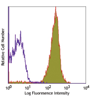
C57BL/6 mouse splenocytes were stained with CD49b/DX5 APC and NK1.1 (clone PK136) Brilliant Violet 605™ (top) or mouse IgG2a, κ Brilliant Violet 605™ isotype control (bottom). -

NK-1.1 surface antigen, also known as CD161b/CD161c and Ly-55, is encoded by the NKR-P1B/NKR-P1C gene. It is expressed on NK cells and NK-T cells in some mouse strains, including C57BL/6, FVB/N, and NZB, but not AKR, BALB/c, CBA/J, C3H, DBA/1, DBA/2, NOD, SJL, and 129. Expression of NKR-P1C antigen has been correlated with lysis of tumor cells in vitro and rejection of bone marrow allografts in vivo. NK-1.1 has also been shown to play a role in NK cell activation, IFN-γ production, and cytotoxic granule release. NK-1.1 and DX5 are commonly used as mouse NK cell markers.
Product DetailsProduct Details
- Reactivity
- Mouse
- Antibody Type
- Monoclonal
- Host Species
- Mouse
- Immunogen
- NK-1+ cells from mouse spleen and bone marrow
- Formulation
- Phosphate-buffered solution, pH 7.2, containing 0.09% sodium azide and BSA (origin USA).
- Preparation
- The antibody was purified by affinity chromatography and conjugated with Brilliant Violet 605™ under optimal conditions.
- Concentration
- µg sizes: 0.2 mg/mLµL sizes: lot-specific (to obtain lot-specific concentration and expiration, please enter the lot number in our Certificate of Analysis online tool.)
- Storage & Handling
- The antibody solution should be stored undiluted between 2°C and 8°C, and protected from prolonged exposure to light. Do not freeze.
- Application
-
FC - Quality tested
- Recommended Usage
-
Each lot of this antibody is quality control tested by immunofluorescent staining with flow cytometric analysis. For flow cytometric staining using the µl sizes, the suggested use of this reagent is 5 µl per million cells in 100 µl staining volume or 5 µl per 100 µl of whole blood. For flow cytometric staining using the µg size, the suggested use of this reagent is ≤0.5 µg per million cells in 100 µl volume. It is recommended that the reagent be titrated for optimal performance for each application.
Brilliant Violet 605™ excites at 405 nm and emits at 603 nm. The bandpass filter 610/20 nm is recommended for detection, although filter optimization may be required depending on other fluorophores used. Be sure to verify that your cytometer configuration and software setup are appropriate for detecting this channel. Refer to your instrument manual or manufacturer for support. Brilliant Violet 605™ is a trademark of Sirigen Group Ltd.
Learn more about Brilliant Violet™.
This product is subject to proprietary rights of Sirigen Inc. and is made and sold under license from Sirigen Inc. The purchase of this product conveys to the buyer a non-transferable right to use the purchased product for research purposes only. This product may not be resold or incorporated in any manner into another product for resale. Any use for therapeutics or diagnostics is strictly prohibited. This product is covered by U.S. Patent(s), pending patent applications and foreign equivalents. - Excitation Laser
-
Violet Laser (405 nm)
- Application Notes
-
Additional reported applications (for the relevant formats) include: immunoprecipitation1,2, complement-dependent cytotoxicity3, in vivo depletion4,5,9,10, mediation of in vitro redirected lysis6, blocking of NK cell function7, induction of proliferation8, immunohistochemical staining of frozen sections11, immunofluorescence microscopy11, and spatial biology (IBEX)16,17. The LEAF™ purified antibody (Endotoxin <0.1 EU/µg, Azide-Free, 0.2 µm filtered) is recommended for functional assays (Cat. No. 108712).
-
Application References
(PubMed link indicates BioLegend citation) -
- Carlyle JR, et al. 1999. J. Immunol. 162:5917. (IP)
- Sentman CL, et al. 1989. Hybridoma 8:605. (IP)
- Koo GC, et al. 1984. Hybridoma 3:301. (Cyt)
- Sentman CL, et al. 1989. J. Immunol. 142:1847. (Deplete)
- Koo GC, et al. 1986. J. Immunol. 137:3742. (Deplete)
- Karlhofer FM, et al. 1991. J. Immunol. 146:3662.
- Kung SK, et al. 1999. J. Immunol. 162:5876. (Block)
- Reichlin A, et al. 1998. Immunol. Cell Biol. 76:143.
- Drobyski W, et al. 1996. Blood 87:5355. (Deplete)
- Andoniou CE, et al. 2005. Nat. Immunol. 6:1011. (Deplete)
- Kanwar JR, et al. 2001. J. Natl. Cancer Inst. 93:1541. (IHC, IF)
- Kroemer A, et al. 2008. J. Immunol. 180:7818. PubMed
- Kim JY, et al. 2009. Exp Mol Med. 30:288. PubMed
- Bankoti J, et al. 2010. Toxicol. Sci. 115:422. (FC) PubMed
- Lee H, et al. 2014. Invest Ophthalmol Vis Sci. 55:2885. PubMed
- Radtke AJ, et al. 2020. Proc Natl Acad Sci U S A. 117:33455-65. (SB) PubMed
- Radtke AJ, et al. 2022. Nat Protoc. 17:378-401. (SB) PubMed
- Product Citations
- RRID
-
AB_2562273 (BioLegend Cat. No. 108739)
AB_2686977 (BioLegend Cat. No. 108753)
AB_2562274 (BioLegend Cat. No. 108740)
Antigen Details
- Structure
- NKR-P1 gene family
- Distribution
-
NK and NK-T cells in the NK1.1 mouse strains (C57BL, FVB/N, NZB)
- Function
- NK cell activation, IFN-γ production, cytotoxic granule release
- Cell Type
- NK cells, NKT cells
- Biology Area
- Immunology, Innate Immunity
- Antigen References
-
1. Lanier LL. 1997. Immunity 6:371.
2. Yokoyama WM, et al. 1993. Ann. Rev. Immunol. 11:613.
3. Koo GC, et al. 1986. J. Immunol. 137:3742.
4. Giorda R, et al. 1991. J. Immunol. 147:1701. - Gene ID
- 17059 View all products for this Gene ID
- UniProt
- View information about NK-1.1 on UniProt.org


Related FAQs
Customers Also Purchased
Compare Data Across All Formats
This data display is provided for general comparisons between formats.
Your actual data may vary due to variations in samples, target cells, instruments and their settings, staining conditions, and other factors.
If you need assistance with selecting the best format contact our expert technical support team.


Follow Us