 Login / Register


- Clone
- SK1 (See other available formats)
- Regulatory Status
- RUO
- Other Names
- T8, Leu2
- Isotype
- Mouse IgG1, κ
- Ave. Rating
- Submit a Review
- Product Citations
- publications

-

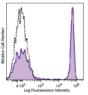
Human peripheral blood lymphocytes were stained with CD8 (clone SK1) Brilliant Violet 785™ (filled histogram) or mouse IgG1, κ Brilliant Violet 785™ isotype control (open histogram).
| Cat # | Size | Price | Quantity Check Availability | Save | ||
|---|---|---|---|---|---|---|
| 344739 | 25 tests | 164€ | ||||
| 344740 | 100 tests | 335€ | ||||
CD8a is a 32-34 kD type I glycoprotein. It forms a homodimer (CD8a/a) or heterodimer (CD8a/b) with CD8b. CD8, also known as T8 and Leu2, is a member of the immunoglobulin superfamily found on the majority of thymocytes, a subset of peripheral blood T cells, and NK cells (which express almost exclusively CD8a homodimers). CD8 acts as a co-receptor with MHC class I-restricted T cell receptors in antigen recognition and T cell activation and has been shown to play a role in thymic differentiation. Two domains in CD8a are important for function: the extracellular IgSF domain binds the α3 domain of MHC class I and the cytoplasmic CXCP motif binds the tyrosine kinase p56 Lck.
Product DetailsProduct Details
- Reactivity
- Human,Cynomolgus,Rhesus
- Antibody Type
- Monoclonal
- Host Species
- Mouse
- Formulation
- Phosphate-buffered solution, pH 7.2, containing 0.09% sodium azide and BSA (origin USA).
- Preparation
- The antibody was purified by affinity chromatography and conjugated with Brilliant Violet 785™ under optimal conditions.
- Concentration
- Lot-specific (to obtain lot-specific concentration and expiration, please enter the lot number in our Certificate of Analysis online tool.)
- Storage & Handling
- The antibody solution should be stored undiluted between 2°C and 8°C, and protected from prolonged exposure to light. Do not freeze.
- Application
-
FC - Quality tested
- Recommended Usage
-
Each lot of this antibody is quality control tested by immunofluorescent staining with flow cytometric analysis. For flow cytometric staining, the suggested use of this reagent is 5 µl per million cells in 100 µl staining volume or 5 µl per 100 µl of whole blood.
Brilliant Violet 785™ excites at 405 nm and emits at 785 nm. The bandpass filter 780/60 nm is recommended for detection, although filter optimization may be required depending on other fluorophores used. Be sure to verify that your cytometer configuration and software setup are appropriate for detecting this channel. Refer to your instrument manual or manufacturer for support. Brilliant Violet 785™ is a trademark of Sirigen Group Ltd.
Learn more about Brilliant Violet™.
This product is subject to proprietary rights of Sirigen Inc. and is made and sold under license from Sirigen Inc. The purchase of this product conveys to the buyer a non-transferable right to use the purchased product for research purposes only. This product may not be resold or incorporated in any manner into another product for resale. Any use for therapeutics or diagnostics is strictly prohibited. This product is covered by U.S. Patent(s), pending patent applications and foreign equivalents. - Excitation Laser
-
Violet Laser (405 nm)
- Application Notes
-
Clone SK1 recognizes the a chain of CD8. Additional reported applications (for the relevant formats) include: proteogenomics8, immunohistochemistry of acetone-fixed frozen tissue sections, and spatial biology (IBEX)9,10. This clone was tested in-house and does not demonstrate utility for formalin-fixed paraffin-embedded (FFPE) human tonsil sections.
-
Application References
(PubMed link indicates BioLegend citation) -
- Ledbetter JA, et al. 1981. J. Exp. Med. 153:310.
- Campanelli R, et al. 2002. Intl. Immunol. 14:39.
- Evans RL, et al. 1981. Immunol. 78:544.
- Wooldridge L, et al. 2005. J. Bio. Chem. 280:27491.
- Ch'el IL, et al. 2011. J Exp Med. 208:633. PubMed
- Carbone A, et al. 1999. Blood 93:2319. (IHC-F)
- Ahmed A, et al. 2001. J. Pathol. 193:383. (IHC)
- Peterson VM, et al. 2017. Nat. Biotechnol. 35:936. (PG)
- Radtke AJ, et al. 2020. Proc Natl Acad Sci USA. 117:33455-33465. (SB) PubMed
- Radtke AJ, et al. 2022. Nat Protoc. 17:378-401. (SB) PubMed
- Product Citations
- RRID
-
AB_2566201 (BioLegend Cat. No. 344739)
AB_2566202 (BioLegend Cat. No. 344740)
Antigen Details
- Structure
- Ig superfamily, homodimer or heterodimer with CD8b, 32-34 kD
- Distribution
-
Majority of thymocytes, T cell subset, NK cells
- Function
- MHC class I co-receptor, thymic differentiation, T cell activation
- Ligand/Receptor
- MHC Class I molecules
- Cell Type
- NK cells, T cells, Thymocytes
- Biology Area
- Immunology
- Molecular Family
- CD Molecules
- Antigen References
-
1. Barclay N, et al. 1993. The Leucocyte Antigen FactsBook. Academic Press Inc. San Diego.
- Gene ID
- 925 View all products for this Gene ID
- UniProt
- View information about CD8 on UniProt.org
Related Pages & Pathways
Pathways


Customers Also Purchased
Compare Data Across All Formats
This data display is provided for general comparisons between formats.
Your actual data may vary due to variations in samples, target cells, instruments and their settings, staining conditions, and other factors.
If you need assistance with selecting the best format contact our expert technical support team.


Follow Us