 Login / Register


- Clone
- 6H6 (See other available formats)
- Regulatory Status
- RUO
- Other Names
- IL-3Rα, IL-3 Receptor alpha
- Isotype
- Mouse IgG1, κ
- Ave. Rating
- Submit a Review
- Product Citations
- publications

-

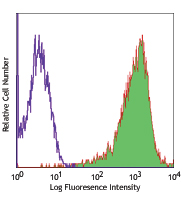
Human peripheral blood lymphocytes stained with 6H6 PE and CD45RO PE/Cy5
| Cat # | Size | Price | Quantity Check Availability | Save | ||
|---|---|---|---|---|---|---|
| 306005 | 25 tests | 66€ | ||||
| 306006 | 100 tests | 140€ | ||||
CD123 is the 70 kD transmembrane α chain of the IL-3 receptor. Alone, CD123 binds IL-3 with low affinity; when CD123 associates with CD131 (common β chain), it binds IL-3 with high affinity. CD123 does not transduce intracellular signals upon binding IL-3 and requires the β chain for this function. CD123 is expressed by myeloid precursors, macrophages, dendritic cells, mast cells, basophils, megakaryocytes, and some B cells.
Product DetailsProduct Details
- Reactivity
- Human
- Antibody Type
- Monoclonal
- Host Species
- Mouse
- Immunogen
- Human IL-3Rα transfected COS cells.
- Formulation
- Phosphate-buffered solution, pH 7.2, containing 0.09% sodium azide and BSA (origin USA)
- Preparation
- The antibody was purified by affinity chromatography, and conjugated with PE under optimal conditions.
- Concentration
- Lot-specific (to obtain lot-specific concentration and expiration, please enter the lot number in our Certificate of Analysis online tool.)
- Storage & Handling
- The antibody solution should be stored undiluted between 2°C and 8°C, and protected from prolonged exposure to light. Do not freeze.
- Application
-
FC - Quality tested
- Recommended Usage
-
Each lot of this antibody is quality control tested by immunofluorescent staining with flow cytometric analysis. For flow cytometric staining, the suggested use of this reagent is 5 µl per million cells in 100 µl staining volume or 5 µl per 100 µl of whole blood.
- Excitation Laser
-
Blue Laser (488 nm)
Green Laser (532 nm)/Yellow-Green Laser (561 nm)
- Application Notes
-
Clone 6H6 does not inhibit IL-3 binding to low- or high-affinity IL-3Rs. Additional reported applications (for the relevant formats) include: Western blotting1, immunoprecipitation1, and immunohistochemical staining of acetone-fixed frozen sections2 and also paraformaldehyde fixed paraffin embedded tissue7.
-
Application References
(PubMed link indicates BioLegend citation) -
- Sun Q, et al. 1996. Blood 87:83. (IP, WB)
- Herling M, et al. 2003. Blood 101:5007. (IHC)
- Charles N, et al. 2010. Nat. Med. 16:701. (FC) PubMed
- Martin-Gayo E, et al. 2010. Blood 115:5366. PubMed
- Chen SC, et al. 2010. Arch Dermatol Res. 302:113. PubMed
- Liu Y, et al. 2012. Food Chem Toxicol. 50:1920. PubMed
- Peduzzi E, et al. 2007. J. Invest. Dermatol. 127:638. (IHC)
- Product Citations
- RRID
-
AB_314579 (BioLegend Cat. No. 306005)
AB_314580 (BioLegend Cat. No. 306006)
Antigen Details
- Structure
- Ig superfamily, type I transmembrane glycoprotein, associates with CDw131, 70 kD
- Distribution
-
Myeloid precursors, basophils, mast cells, macrophages, dendritic cells, megakaryocytes, subset of lymphocytes
- Function
- Hematopoietic cell proliferation, differentiation
- Ligand/Receptor
- IL-3
- Cell Type
- Basophils, Dendritic cells, Hematopoietic stem and progenitors, Lymphocytes, Macrophages, Mast cells, Megakaryocytes
- Biology Area
- Immunology
- Molecular Family
- CD Molecules, Cytokine/Chemokine Receptors
- Antigen References
-
1. Miyajima A, et al. 1993. Blood 82:1960.
- Gene ID
- 3563 View all products for this Gene ID
- UniProt
- View information about CD123 on UniProt.org
Related Pages & Pathways
Pathways


Related FAQs
- What type of PE do you use in your conjugates?
- We use R-PE in our conjugates.
Customers Also Purchased

Compare Data Across All Formats
This data display is provided for general comparisons between formats.
Your actual data may vary due to variations in samples, target cells, instruments and their settings, staining conditions, and other factors.
If you need assistance with selecting the best format contact our expert technical support team.


Follow Us