 Login / Register


- Clone
- 29E.2A3 (See other available formats)
- Regulatory Status
- RUO
- Other Names
- Programmed cell death ligand 1 (PD-L1), B7 homolog 1 (B7-H1)
- Isotype
- Mouse IgG2b, κ
- Ave. Rating
- Submit a Review
- Product Citations
- publications

-

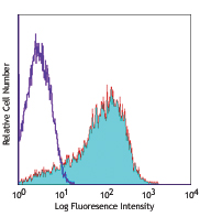
PHA-stimulated (3 days) human peripheral blood lymphocytes were stained with CD274 (clone 29E.2A3) PE (filled histogram) or mouse IgG2b, κ PE isotype control (open histogram).
| Cat # | Size | Price | Quantity Check Availability | Save | ||
|---|---|---|---|---|---|---|
| 329705 | 25 tests | 95€ | ||||
| 329706 | 100 tests | 211€ | ||||
CD274, also known as PD-L1 and B7-H1, is type I transmembrane glycoprotein that serves as a ligand for CD279 (PD-1). This interaction is believed to regulate the balance between the stimulatory and inhibitory signals needed for responses to microbes and maintenance of self-tolerance. CD274 is involved in the costimulation of T cell proliferation and IL-10 and IFN-γ production in an IL-2-dependent and CD279-independent manner. Conflicting data has shown that CD274 can inhibit T cell proliferation and cytokine production, and alternatively, enhance T cell activation. Other studies suggest that CD274 may signal bidirectionally, raising interesting implications for its expression in a wide variety of cell types, including T and B cells, antigen-presenting cells, and nonhematopoietic cells.
Product DetailsProduct Details
- Reactivity
- Human
- Antibody Type
- Monoclonal
- Host Species
- Mouse
- Immunogen
- Full length human PD-L1
- Formulation
- Phosphate-buffered solution, pH 7.2, containing 0.09% sodium azide and BSA (origin USA)
- Preparation
- The antibody was purified by affinity chromatography, and conjugated with PE under optimal conditions.
- Concentration
- Lot-specific (to obtain lot-specific concentration and expiration, please enter the lot number in our Certificate of Analysis online tool.)
- Storage & Handling
- The antibody solution should be stored undiluted between 2°C and 8°C, and protected from prolonged exposure to light. Do not freeze.
- Application
-
FC - Quality tested
- Recommended Usage
-
Each lot of this antibody is quality control tested by immunofluorescent staining with flow cytometric analysis. For flow cytometric staining, the suggested use of this reagent is 5 µl per million cells in 100 µl staining volume or 5 µl per 100 µl of whole blood.
- Excitation Laser
-
Blue Laser (488 nm)
Green Laser (532 nm)/Yellow-Green Laser (561 nm)
- Application Notes
-
Clone 29E.2A3 is reported to recognize an epitope on PD-L1 within the PD-L1-CD80 binding region5. Additional reported applications (for the relevant formats) include: blocking1-3 and immunohistochemical staining of acetone-fixed frozen sections1. The Ultra-LEAF™ purified antibody (Endotoxin < 0.01 EU/µg, Azide-Free, 0.2 µm filtered) is recommended for functional assays (Cat. No. 329715, 329716, 329745 - 329748).
It has been observed that clone 29E.2A3 is able to bind to Alexa Fluor® 700 antibody conjugates during multi-color immunofluorescent staining. This interaction can be resolved by sequentially staining with the 29E.2A3 antibody first and then followed by the Alexa Fluor® 700 conjugate of interest.Clone 29E.2A3 does not work in Western blot applications7.
-
Application References
(PubMed link indicates BioLegend citation) -
- Brown J, et al. 2003. J. Immunol. 170:1257. (FC, IHC, Block)
- Radziewicz H, et al. 2007. J. Virol. 81:2545. (Block)
- Nakamoto N, et al. 2009. PLoS Pathog. 5:e1000313. (Block)
- Barsoum IB, et al. 2014. Cancer Res. 74:665. PubMed
- Haile, S et al. 2013. J. Immunol. 191:2829.
- RL M, et al. 2015. PNAS. 112:6506-6514. PubMed
- Mahoney KM, et al. 2015. Cancer Immunol. Res. 3:1308.
- Product Citations
- RRID
-
AB_940366 (BioLegend Cat. No. 329705)
AB_940368 (BioLegend Cat. No. 329706)
Antigen Details
- Distribution
-
T cells, B cells, NK cells, monocytes/macrophages, granulocytes and dendritic cells
- Function
- CD274 is involved in the costimulatory signal, essential for T lymphocyte proliferation and production of IL-10 and IFN-γ, in an IL-2-dependent and a PD-1-CD1-independent manner. Its interaction with PD-1-CD1 inhibits T-cell proliferation and cytokine production.
- Ligand/Receptor
- PD-1 (PDCD1)
- Cell Type
- B cells, Dendritic cells, Fibroblasts, Granulocytes, Macrophages, Monocytes, NK cells, T cells
- Biology Area
- Cancer Biomarkers, Costimulatory Molecules, Immunology
- Molecular Family
- Adhesion Molecules, CD Molecules, Immune Checkpoint Receptors
- Antigen References
-
1. Sharpe A, et al. 2007. Nat. Immunol. 8:239.
- Gene ID
- 29126 View all products for this Gene ID
- UniProt
- View information about CD274 on UniProt.org


Related FAQs
- What type of PE do you use in your conjugates?
- We use R-PE in our conjugates.
Customers Also Purchased
Compare Data Across All Formats
This data display is provided for general comparisons between formats.
Your actual data may vary due to variations in samples, target cells, instruments and their settings, staining conditions, and other factors.
If you need assistance with selecting the best format contact our expert technical support team.


Follow Us