 Login / Register


- Clone
- A161A1 (See other available formats)
- Regulatory Status
- RUO
- Other Names
- T4/Leu-3
- Isotype
- Rat IgG2b, κ
- Ave. Rating
- Submit a Review
- Product Citations
- publications

-

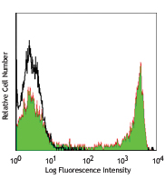
Human peripheral blood lymphocytes were stained with CD4 (clone A161A1) PE (filled histogram) or rat IgG2b, κ PE isotype control (open histogram).
| Cat # | Size | Price | Quantity Check Availability | Save | ||
|---|---|---|---|---|---|---|
| 357403 | 25 tests | 59€ | ||||
| 357404 | 100 tests | 128€ | ||||
CD4, also known as T4/Leu-3, is a 55 kD single-chain type I transmembrane glycoprotein and member of the immunoglobulin superfamily. It is expressed on most thymocytes, helper T cells, type II NKT cells, and monocytes/macrophages. CD4 is part of the TCR/CD3 complex, binds to β2 domain from the MHC class II molecule, and participates in TCR signal transduction. CD4 is the receptor of IL-16 and is a coreceptor for the human immunodeficiency virus (HIV) and human herpes virus 7 (HHV-7).
Product DetailsProduct Details
- Reactivity
- Human
- Antibody Type
- Monoclonal
- Host Species
- Rat
- Immunogen
- Human CD4 T cells
- Formulation
- Phosphate-buffered solution, pH 7.2, containing 0.09% sodium azide and BSA (origin USA)
- Preparation
- The antibody was purified by affinity chromatography and conjugated with PE under optimal conditions.
- Concentration
- Lot-specific (to obtain lot-specific concentration and expiration, please enter the lot number in our Certificate of Analysis online tool.)
- Storage & Handling
- The antibody solution should be stored undiluted between 2°C and 8°C, and protected from prolonged exposure to light. Do not freeze.
- Application
-
FC - Quality tested
- Recommended Usage
-
Each lot of this antibody is quality control tested by immunofluorescent staining with flow cytometric analysis. For flow cytometric staining, the suggested use of this reagent is 5 µl per million cells in 100 µl staining volume or 5 µl per 100 µl of whole blood.
- Excitation Laser
-
Blue Laser (488 nm)
Green Laser (532 nm)/Yellow-Green Laser (561 nm)
- Product Citations
- RRID
-
AB_2562035 (BioLegend Cat. No. 357403)
AB_2562036 (BioLegend Cat. No. 357404)
Antigen Details
- Structure
- 55 kD, type I transmembrane glycoprotein, member of the Ig superfamily
- Distribution
-
Helper T cells, subset of thymocytes, monocytes/macrophages, type II NKT cells
- Interaction
- Part of the TCR/CD3 complex
- Ligand/Receptor
- β2 domain from the MHC class II
- Cell Type
- T cells, Monocytes, Macrophages, NKT cells
- Biology Area
- Immunology
- Molecular Family
- CD Molecules
- Antigen References
-
1. Zhu J, et al. 2010. Annu Rev. Immunol. 28:445.
2. Vignali DA. 2010. J. Immunol. 184:5933.
3. Zhou L, et al. 2009. Immunity 30:646.
4. Singer A, et al. 2008. Nat. Rev. Immunol. 8:788.
5. Zhu J and Paul WE. 2008. Blood 112:1557. - Gene ID
- 920 View all products for this Gene ID
- UniProt
- View information about CD4 on UniProt.org
Related Pages & Pathways
Pathways


Related FAQs
- I am unable to see expression of T cell markers such as CD3 and CD4 post activation.
- TCR-CD3 complexes on the T-lymphocyte surface are rapidly downregulated upon activation with peptide-MHC complex, superantigen or cross-linking with anti-TCR or anti-CD3 antibodies. PMA/Ionomycin treatment has been shown to downregulate surface CD4 expression. Receptor downregulation is a common biological phenomenon and so make sure that your stimulation treatment is not causing it in your sample type.
- What type of PE do you use in your conjugates?
- We use R-PE in our conjugates.
Customers Also Purchased
Compare Data Across All Formats
This data display is provided for general comparisons between formats.
Your actual data may vary due to variations in samples, target cells, instruments and their settings, staining conditions, and other factors.
If you need assistance with selecting the best format contact our expert technical support team.


Follow Us