 Login / Register


- Clone
- FN50 (See other available formats)
- Regulatory Status
- RUO
- Workshop
- IV A91
- Other Names
- Very Early Activation Antigen (VEA), Activation inducer molecule (AIM)
- Isotype
- Mouse IgG1, κ
- Ave. Rating
- Submit a Review
- Product Citations
- publications

-

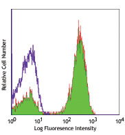
PMA + ionomycin stimulated (6 hours) human lymphocytes stained with FN50 PE
| Cat # | Size | Price | Quantity Check Availability | Save | ||
|---|---|---|---|---|---|---|
| 310905 | 25 tests | 53€ | ||||
| 310906 | 100 tests | 115€ | ||||
CD69 is a 27-33 kD type II transmembrane protein also known as activation inducer molecule (AIM), very early activation antigen (VEA), and MLR3. It is a member of the C-type lectin family, expressed as a disulfide-linked homodimer. Other members of this receptor family include NKG2, NKR-P1 CD94, and Ly49. CD69 is transiently expressed on activated leukocytes including T cells, thymocytes, B cells, NK cells, neutrophils, and eosinophils. CD69 is constitutively expressed by a subset of medullary mature thymocytes, platelets, mantle B cells, and certain CD4+ T cells in germinal centers of normal lymph nodes. CD69 is involved in early events of lymphocyte, monocyte, and platelet activation, and has a functional role in redirected lysis mediated by activated NK cells.
Product DetailsProduct Details
- Reactivity
- Human
- Antibody Type
- Monoclonal
- Host Species
- Mouse
- Formulation
- Phosphate-buffered solution, pH 7.2, containing 0.09% sodium azide and BSA (origin USA)
- Preparation
- The antibody was purified by affinity chromatography, and conjugated with PE under optimal conditions.
- Concentration
- Lot-specific (to obtain lot-specific concentration and expiration, please enter the lot number in our Certificate of Analysis online tool.)
- Storage & Handling
- The CD69 antibody solution should be stored undiluted between 2°C and 8°C, and protected from prolonged exposure to light. Do not freeze.
- Application
-
FC - Quality tested
- Recommended Usage
-
Each lot of this antibody is quality control tested by immunofluorescent staining with flow cytometric analysis. For flow cytometric staining, the suggested use of this reagent is 5 µl per million cells in 100 µl staining volume or 5 µl per 100 µl of whole blood.
- Excitation Laser
-
Blue Laser (488 nm)
Green Laser (532 nm)/Yellow-Green Laser (561 nm)
- Application Notes
-
Additional reported applications (for the relevant formats) include: immunohistochemical staining of acetone-fixed frozen tissue sections2, immunofluorescence microscopy3, and spatial biology (IBEX)8,9.
-
Application References
(PubMed link indicates BioLegend citation) -
- Knapp WB, et al. 1989. Leucocyte Typing IV. Oxford University Press. New York.
- Sakkas LI, et al. 1998. Clin. and Diag. Lab. Immunol. 5:430. (IHC)
- Kim JR, et al. 2005. BMC Immunol. 6:3. (IF)
- Verjans GM, et al. 2007. P. Natl. Acad. Sci. USA 104:3496.
- Lu H, et al. 2009. Toxicol Sci. 112:363. (FC) PubMed
- Thakral D, et al. 2008. J. Immunol. 180:7431. (FC) PubMed
- Yoshino N, et al. 2000. Exp. Anim. (Tokyo) 49:97. (FC)
- Radtke AJ, et al. 2020. Proc Natl Acad Sci USA. 117:33455-33465. (SB) PubMed
- Radtke AJ, et al. 2022. Nat Protoc. 17:378-401. (SB) PubMed
- Product Citations
- RRID
-
AB_314840 (BioLegend Cat. No. 310905)
AB_314841 (BioLegend Cat. No. 310906)
Antigen Details
- Structure
- C-type lectin, type II glycoprotein, 28/32 kD
- Distribution
-
Activated T cells, B cells, NK cells, granulocytes, thymocytes, platelets, Langerhans cells
- Function
- Lymphocyte, monocyte, and platelet activation, NK cell killing
- Cell Type
- B cells, Granulocytes, Langerhans cells, NK cells, Platelets, T cells, Thymocytes, Tregs
- Biology Area
- Costimulatory Molecules, Immunology
- Molecular Family
- CD Molecules
- Antigen References
-
1. Schlossman S, et al. Eds. 1995. Leucocyte Typing V. Oxford University Press. New York.
2. Testi R, et al. 1994. Immunol. Today 15:479. - Gene ID
- 969 View all products for this Gene ID
- UniProt
- View information about CD69 on UniProt.org
Related Pages & Pathways
Pathways


Related FAQs
- What type of PE do you use in your conjugates?
- We use R-PE in our conjugates.
Customers Also Purchased
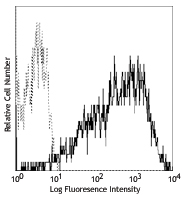
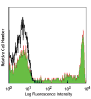
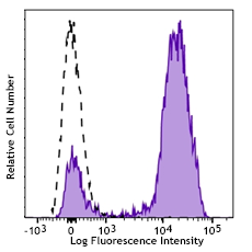
Compare Data Across All Formats
This data display is provided for general comparisons between formats.
Your actual data may vary due to variations in samples, target cells, instruments and their settings, staining conditions, and other factors.
If you need assistance with selecting the best format contact our expert technical support team.


Follow Us