 Login / Register


- Clone
- 2D10 (See other available formats)
- Regulatory Status
- RUO
- Workshop
- VI CD80.1
- Other Names
- B7-1, B7, BB1
- Isotype
- Mouse IgG1, κ
- Ave. Rating
- Submit a Review
- Product Citations
- publications

-

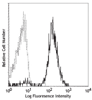
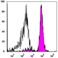
Human B-cell Burkitt's lymphoma cell line Daudi stained with 2D10 PE
| Cat # | Size | Price | Quantity Check Availability | Save | ||
|---|---|---|---|---|---|---|
| 305207 | 25 tests | 80€ | ||||
| 305208 | 100 tests | 169€ | ||||
CD80, also known as B7-1, B7, and BB1, is a 60 kD single chain type I glycoprotein belonging to the immunoglobulin superfamily. CD80 is expressed on activated B and T cells, macrophages, and dendritic cells. CD80 binds to CD28 and CD152 (CTLA-4). Along with CD86, CD80 plays a critical role in regulation of T cell activation. The interaction of CD80 with CD28 provides a potent costimulatory signal for T cell activation through the CD3 complex, while its interaction with CTLA-4 provides an inhibitory signal for T cell activation.
Product DetailsProduct Details
- Reactivity
- Human
- Antibody Type
- Monoclonal
- Host Species
- Mouse
- Formulation
- Phosphate-buffered solution, pH 7.2, containing 0.09% sodium azide and BSA (origin USA)
- Preparation
- The antibody was purified by affinity chromatography, and conjugated with PE under optimal conditions.
- Concentration
- Lot-specific (to obtain lot-specific concentration and expiration, please enter the lot number in our Certificate of Analysis online tool.)
- Storage & Handling
- The antibody solution should be stored undiluted between 2°C and 8°C, and protected from prolonged exposure to light. Do not freeze.
- Application
-
FC - Quality tested
- Recommended Usage
-
Each lot of this antibody is quality control tested by immunofluorescent staining with flow cytometric analysis. For flow cytometric staining, the suggested use of this reagent is 5 µl per million cells in 100 µl staining volume or 5 µl per 100 µl of whole blood.
- Excitation Laser
-
Blue Laser (488 nm)
Green Laser (532 nm)/Yellow-Green Laser (561 nm)
- Application Notes
-
Additional reported applications (for the relevant formats) include: in vitro blocking of T cell activation, immunohistochemical staining of acetone-fixed frozen tissue sections2, immunoprecipitation, and Western blotting3. The Ultra-LEAF™ purified antibody (Endotoxin <0.1 EU/µg, Azide-Free, 0.2 µm filtered) is recommended for functional assays (Cat. Nos. 305245 & 305246).
-
Application References
(PubMed link indicates BioLegend citation) -
- Kishimoto T, et al. Eds. 1997. Leucocyte Typing VI. Garland Publishing Inc. London.
- Battifora M. 1998. J. Clin. Endocr. Metab. 83:4130. (IHC)
- Van der Merwe PA, et al. 1997. J. Exp. Med. 185:3. (WB)
- Jayakumar A, et al. 2008. Infect. Immun. 76:2138. PubMed
- Schubert DA, et al. 2012. J. Exp Med. 209:335. PubMed
- Wen T, et al. 2014. J Immunol. 192:5481. PubMed
- Product Citations
- RRID
-
AB_314503 (BioLegend Cat. No. 305207)
AB_314504 (BioLegend Cat. No. 305208)
Antigen Details
- Structure
- Ig superfamily, type I transmembrane glycoprotein, 60 kD
- Distribution
-
Activated B cells and T cells, macrophages, and dendritic cells
- Function
- T cell costimulation
- Ligand/Receptor
- CD28, CD152 (CTLA-4)
- Cell Type
- B cells, Dendritic cells, Macrophages, T cells, Tregs
- Biology Area
- Cell Biology, Costimulatory Molecules, Immunology, Neuroscience, Neuroscience Cell Markers
- Molecular Family
- CD Molecules, Immune Checkpoint Receptors
- Antigen References
-
1. Freeman G, et al. 1991. J. Exp. Med. 174:625.
2. Linsley P, et al. 1996. Immunity 4:535.
3. Linsley P, et al. 1991. J. Exp. Med. 174:561. - Gene ID
- 941 View all products for this Gene ID
- UniProt
- View information about CD80 on UniProt.org
Related Pages & Pathways
Pathways


Related FAQs
- What type of PE do you use in your conjugates?
- We use R-PE in our conjugates.
Customers Also Purchased
Compare Data Across All Formats
This data display is provided for general comparisons between formats.
Your actual data may vary due to variations in samples, target cells, instruments and their settings, staining conditions, and other factors.
If you need assistance with selecting the best format contact our expert technical support team.


Follow Us