 Login / Register


- Clone
- 29F.1A12 (See other available formats)
- Regulatory Status
- RUO
- Other Names
- PD-1, Programmed Death-1, PDCD1
- Isotype
- Rat IgG2a, κ
- Ave. Rating
- Submit a Review
- Product Citations
- publications

-

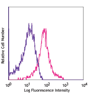
Con-A and IL-2 stimulated (3 days) C57BL/6 splenocytes stained with 29F.1A12 PE and CD3 (145-2C11) APC -

Con A stimulated (3 days) C57BL/6 splenocytes stained with rat IgG2a isotype PE and CD3e (145-2C11) APC
| Cat # | Size | Price | Quantity Check Availability | Save | ||
|---|---|---|---|---|---|---|
| 135205 | 50 µg | 96€ | ||||
| 135206 | 200 µg | 248€ | ||||
CD279, also known as programmed death-1 (PD-1), is a 50-55 kD glycoprotein belonging to the CD28 family of the Ig superfamily. PD-1 is expressed on activated splenic T and B cells and thymocytes. It is induced on activated myeloid cells as well. PD-1 is involved in lymphocyte clonal selection and peripheral tolerance through binding its ligands, B7-H1 (PD-L1) and B7-DC (PD-L2). It has been reported that PD-1 and PD-L1 interactions are critical to positive selection and play a role in shaping the T cell repertoire. PD-L1 negative costimulation is essential for prolonged survival of intratesticular islet allografts.
Product DetailsProduct Details
- Reactivity
- Mouse
- Antibody Type
- Monoclonal
- Host Species
- Rat
- Immunogen
- PD-1 cDNA followed by PD-1-Ig fusion protein
- Formulation
- Phosphate-buffered solution, pH 7.2, containing 0.09% sodium azide.
- Preparation
- The antibody was purified by affinity chromatography, and conjugated with PE under optimal conditions.
- Concentration
- 0.2 mg/ml
- Storage & Handling
- The antibody solution should be stored undiluted between 2°C and 8°C, and protected from prolonged exposure to light. Do not freeze.
- Application
-
FC - Quality tested
- Recommended Usage
-
Each lot of this antibody is quality control tested by immunofluorescent staining with flow cytometric analysis. For flow cytometric staining, the suggested use of this reagent is ≤1.0 µg per million cells in 100 µl volume. It is recommended that the reagent be titrated for optimal performance for each application.
- Excitation Laser
-
Blue Laser (488 nm)
Green Laser (532 nm)/Yellow-Green Laser (561 nm)
- Application Notes
-
Additional reported applications (for the relevant formats) include: immunohistochemical staining of acetone-fixed frozen tissue3, in vivo blocking of PD-1 binding to its ligands2,3, and spatial biology (IBEX)5,6.
-
Application References
(PubMed link indicates BioLegend citation) -
- Good-Jacobson KL, et al. 2010. Nat. Immunol. 11:535. (FC) PubMed
- Lázár-Molnár E, et al. 2008. Proc. Natl. Acad. Sci. USA 105:2658. (Block)
- Liang SC, et al. 2003. Eur. J. Immunol. 33:2706. (FC, IHC, Block)
- Tobias J, et al. 2020. Front Immunol. 11:895 (FC, ELISA) PubMed
- Radtke AJ, et al. 2020. Proc Natl Acad Sci U S A. 117:33455-65. (SB) PubMed
- Radtke AJ, et al. 2022. Nat Protoc. 17:378-401. (SB) PubMed
- Product Citations
- RRID
-
AB_1877232 (BioLegend Cat. No. 135205)
AB_1877231 (BioLegend Cat. No. 135206)
Antigen Details
- Structure
- A 50-55 kD glycoprotein belonging to the CD28 family of the Ig superfamily.
- Distribution
-
Induced on splenic T and B lymphocytes, thymocytes, and myeloid cells after stimulation.
- Function
- Involved in lymphocyte clonal selection and peripheral tolerance, prolonged survival of allografts.
- Ligand/Receptor
- B7-H1 (PD-L1) and B7-DC (PD-L2)
- Cell Type
- B cells, T cells
- Biology Area
- Cancer Biomarkers, Immunology, Inhibitory Molecules
- Molecular Family
- CD Molecules, Immune Checkpoint Receptors
- Antigen References
-
1. Nishimura H, et al. 2001. Science 291:319
2. Agata Y, et al. 1996. Int. Immunol. 8:765
3. Liang SC, et al. 2003. Eur. J. Immunol. 33:2706
4. Barber DL, et al. 2006. Nature 439:682
5. Keir ME, et al. 2005. J. Immunol. 175:7372
6. Koehn BH. et al. 2008. J Immunol. 181:5313 - Gene ID
- 18566 View all products for this Gene ID
- UniProt
- View information about CD279 on UniProt.org
Related Pages & Pathways
Pathways


Related FAQs
- What type of PE do you use in your conjugates?
- We use R-PE in our conjugates.
Customers Also Purchased
Compare Data Across All Formats
This data display is provided for general comparisons between formats.
Your actual data may vary due to variations in samples, target cells, instruments and their settings, staining conditions, and other factors.
If you need assistance with selecting the best format contact our expert technical support team.


Follow Us