PE/Cyanine7 anti-mouse IFN-γ Antibody

Pricing & Availability
Clone
XMG1.2 (See other available formats)
Regulatory Status
RUO
Other Names
Interferon-γ, Immune interferon, Type II interferon, T cell interferon, Macrophage-activating factor (MAF)
Isotype
Rat IgG1, κ
Ave. Rating
Submit a Review
Product Citations
publications
XMG1-2_PECyanine7_IFN-y_Antibody_1_041124
PMA/Ionomycin-stimulated (6hrs) C57BL/6 mouse splenocytes stained with B220 (RA3-6B2) APC and XMG1.2 PE/Cyanine7
  • XMG1-2_PECyanine7_IFN-y_Antibody_1_041124
    PMA/Ionomycin-stimulated (6hrs) C57BL/6 mouse splenocytes stained with B220 (RA3-6B2) APC and XMG1.2 PE/Cyanine7
See PE/Cyanine7 spectral data
Cat # Size Price Quantity Check Availability Save
505825 25 µg 106€
Check Availability


Need larger quantities of this item?
Request Bulk Quote
505826 100 µg 243€
Check Availability


Need larger quantities of this item?
Request Bulk Quote
Description

IFN-γ is a potent multifunctional cytokine which is secreted primarily by activated NK cells and T cells. Originally characterized based on anti-viral activities, IFN-γ also exerts anti-proliferative, immunoregulatory, and proinflammatory activities. IFN-γ can upregulate MHC class I and II antigen expression by antigen-presenting cells.

Product Details
Technical data sheet

Product Details

Reactivity
Mouse
Antibody Type
Monoclonal
Host Species
Rat
Immunogen
E. coli-expressed, recombinant mouse IFN-γ
Formulation
Phosphate-buffered solution, pH 7.2, containing 0.09% sodium azide.
Preparation
The antibody was purified by affinity chromatography, and conjugated with PE/Cyanine7 under optimal conditions.
Concentration
0.2 mg/ml
Storage & Handling
The antibody solution should be stored undiluted between 2°C and 8°C, and protected from prolonged exposure to light. Do not freeze.
Application

ICFC - Quality tested

Recommended Usage

Each lot of this antibody is quality control tested by intracellular immunofluorescent staining with flow cytometric analysis. For flow cytometric staining, the suggested use of this reagent is ≤1.0 µg per million cells in 100 µl volume. It is recommended that the reagent be titrated for optimal performance for each application.

Excitation Laser
Blue Laser (488 nm)
Green Laser (532 nm)/Yellow-Green Laser (561 nm)
Application Notes

ELISA1-4,11,14 or ELISPOT5 Detection: The biotinylated XMG1.2 antibody is useful as a detection antibody for a sandwich ELISA or ELISPOT assay, when used in conjunction with purified R4-6A2 antibody (Cat. No. 505702/505706) as the capture antibody and recombinant mouse IFN-γ (Cat. No. 575309) as the standard.
ELISA or ELISPOT Capture: The purified XMG1.2 antibody is useful as a capture antibody for a sandwich ELISA or ELISPOT assay, when used in conjunction with biotinylated R4-6A2 antibody (Cat. No. 505704) as the detection antibody and recombinant mouse IFN-γ (Cat. No. 575309) as the standard. The LEAF™ purified antibody is suggested for ELISPOT capture (Cat. No. 505812).
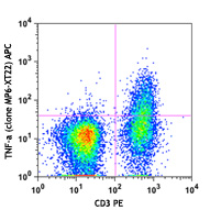
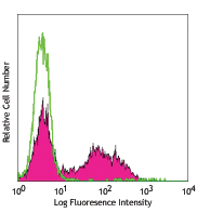
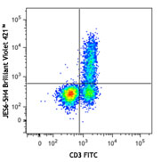
Flow Cytometry7,8,12,13,16: The fluorochrome-labeled XMG1.2 antibody is useful for intracellular immunofluorescent staining and flow cytometric analysis to identify IFN-γ-producing cells within mixed cell populations.
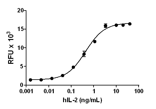
Neutralization1-3,9,10: The XMG1.2 antibody can neutralize the bioactivity of natural or recombinant IFN-γ. The LEAF™ purified antibody (Endotoxin <0.1 EU/µg, Azide-Free, 0.2 µm filtered) is recommended for neutralization of mouse IFN-γ bioactivity in vivo and in vitro (Cat. No. 505812). For in vivo studies or highly sensitive assays, we recommend Ultra-LEAF™ purified antibody (Cat. No. 505834) with a lower endotoxin limit than standard LEAF™ purified antibodies (Endotoxin <0.01 EU/µg).
Additional reported applications (for the relevant formats) include: Western blotting, immunohistochemical staining of frozen tissue sections6,22,23, and immunocytochemistry.
Note: For testing mouse IFN-γ in serum, plasma or supernatant, BioLegend's ELISA Max™ Sets (Cat. No. 430801 to 430806) are specially developed and recommended.

Additional Product Notes

BioLegend is in the process of converting the name PE/Cy7 to PE/Cyanine7. The dye molecule remains the same, so you should expect the same quality and performance from our PE/Cyanine7 products. Please contact Technical Service if you have any questions.

Application References

(PubMed link indicates BioLegend citation)
  1. Abrams J, et al. 1992. Immunol. Rev. 127:5. (ELISA, Neut)
  2. Sander B, et al. 1993. J. Immunol. Meth. 166:201. (ELISA, Neut)
  3. Abrams J, et al. 1995. Curr. Prot. Immunol. John Wiley and Sons, New York. Unit 6.20. (ELISA, Neut)
  4. Yang X, et al. 1993. J. Immunoassay 14:129. (ELISA)
  5. Klinman D, et al. 1994. Curr. Prot. Immunol. John Wiley and Sons, New York. Unit 6.19. (ELISPOT)
  6. Sander B, et al. 1991. Immunol. Rev. 119:65. (IHC)
  7. Ferrick D, et al. 1995. Nature 373:255. (FC)
  8. Ko SY, et al. 2005. J. Immunol. 175:3309. (FC) PubMed
  9. Peterson KE, et al. 2000. J. Virol. 74:5363. (Neut)
  10. DeKrey GK, et al. 1998. Infect. Immun. 66:827. (Neut)
  11. Dzhagalov I, et al. 2007. J. Immunol. 178:2113. (ELISA)
  12. Lawson BR, et al. 2007. J. Immunol. 178:5366. (FC)
  13. Lee JW, et al. 2006. Nature Immunol. 8:181. (FC) PubMed
  14. Xu G, et al. 2007. J. Immunol. 179:5358. (ELISA) PubMed
  15. Montfort M, et al.2004. J. Immunol. 173:4084. PubMed
  16. Haring JS, et al. 2008. J. Immunol. 180:2855. (FC) PubMed
  17. Jordan JM, et al. 2008. Infect Immun. 76:3717. PubMed
  18. Tonkin DR, et al. 2008. J. Immunol. 181:4516. PubMed
  19. Charles N, et al. 2010. Nat. Med. 16:701. (FC) PubMed
  20. Cui Y, et al. 2009. Invest. Ophth. Vis. Sci. 50:5811. (FC) PubMed
  21. Mykkanen OT, et al. 2014. PLoS One. 9:114790. PubMed
  22. Yokogawa M, et al. 2013. Mol. Carcinog. 52:760. (IHC)
  23. Mottram PL, et al. 1998. J Immunol. 161:602. (IHC)
Product Citations
  1. Sun X, et al. 2017. Microbes Infect. 10.1016/j.micinf.2017.08.007. PubMed
  2. Ma W, et al. 2017. Sci Rep. . 10.1038/s41598-017-15661-6. PubMed
  3. Saigusa R, et al. 2018. Arthritis Res Ther. 20:23. PubMed
  4. Habib S, et al. 2018. Infect Immun. 86:e00019. PubMed
  5. Abhyankar MM, et al. 2018. NPJ Vaccines. 3:22. PubMed
  6. Xi‐Zhi J Guo et al. 2018. Immunity. 49(3):531-544 . PubMed
  7. Álvaro de Mingo Pulido et al. 2018. Cancer cell. 33(1):60-74 . PubMed
  8. Lino AC et al. 2018. Immunity. 49(1):120-133 . PubMed
  9. Colliou N, et al. 2018. Gut Microbes. 9:279. PubMed
  10. Nagai Y, et al. 2019. Front Immunol. 10:174. PubMed
  11. Kosack L, et al. 2017. Sci Rep. 7:11289. PubMed
  12. Laura C Burzynski et al. 2019. Immunity. 50(4):1033-1042 . PubMed
  13. Imbratta C, et al. 2019. Sci Rep. 9:6135. PubMed
  14. Choi H et al. 2019. Cell Rep. 27(3):806-819 . PubMed
  15. Li C, et al. 2018. Cell. 174:285. PubMed
  16. Findlay EG, et al. 2019. Oncoimmunology. 8:1608106. PubMed
  17. Omenetti S, et al. 2019. Immunity. 51:77. PubMed
  18. Britton GJ et al. 2019. Immunity. 50(1):212-224 . PubMed
  19. Resende M, et al. 2019. J Immunol. 203:2451. PubMed
  20. Wu J et al. 2019. Immunity. 50(5):1218-1231 . PubMed
  21. Christopher S Garris et al. 2018. Immunity. 49(6):1148-1161 . PubMed
  22. Oliveira AC et al. 2017. eLife. 6 pii: e30883. PubMed
  23. Overacre-Delgoffe AE, et al. 2017. Cell. 169:1130. PubMed
  24. Deng Z, et al. 2017. Oncogene. 36:639. PubMed
  25. Liu QZ, et al. 2018. Front Immunol. 1.131944444. PubMed
  26. Wang X, et al. 2019. Cell Res. 29:787. PubMed
  27. Wolf Y, et al. 2019. Cell. 179:219. PubMed
  28. Lercher A, et al. 2019. Immunity. 51:1074. PubMed
  29. Fu S, et al. 2020. Nat Commun. 0.7625. PubMed
  30. Jing Y, et al. 2020. Sci Adv. 6:eaax9455. PubMed
  31. Zhang W, et al. 2020. Nat Commun. 11:1187. PubMed
  32. Maluski M, et al. 2019. J Clin Invest. 129:5108. PubMed
  33. Schadt L, et al. 2020. Cell Reports. 29(5):1236-1248.e7.. PubMed
  34. Zhao B, et al. 2020. Nat Commun. 11:908. PubMed
  35. Wu B, et al. 2020. Int J Biol Sci. 16:1526. PubMed
  36. Lin H, et al. 2020. Front Microbiol. 1.330555556. PubMed
  37. Nakata J, et al. 2018. Oncotarget. 9:36029. PubMed
  38. Zander R, et al. 2020. Immunity. 51(6):1028-1042.e4.. PubMed
  39. Zhang H, et al. 2020. Cancer Cell. 37(1):37-54.e9.. PubMed
  40. Harel M, et al. 2020. Cell. 179(1):236-250.e18.. PubMed
  41. Wu J, et al. 2020. Cell Reports. 31(1):107484. PubMed
  42. He Y, et al. 2020. Sci Adv. 6:eaba7589. PubMed
  43. Wu J, et al. 2020. Sci Adv. 6:eaba3458. PubMed
  44. Lee J, et al. 2007. Nat Immunol. 8:181. PubMed
  45. Sawant D, et al. 2014. J Immunol. 192:2904. PubMed
  46. Friedman R, et al. 2014. Proc Natl Acad Sci U S A. 111:9223. PubMed
  47. Madireddi S, et al. 2014. J Exp Med. 211:1433. PubMed
  48. Shiomi A, et al. 2014. J Immunol. 193:849. PubMed
  49. Burrack K, et al. 2015. J Immunol. 194:678. PubMed
  50. Holm J, et al. 2015. PLoS One. 10:125495. PubMed
  51. Wen J, et al. 2015. J Pharmacol Sci. 128: 116-124. PubMed
  52. Olguín J, et al. 2015. Microbes Infect. 17: 586-595. PubMed
  53. Glennie N, et al. 2015. J Exp Med. 212: 1405-1414. PubMed
  54. Meadows D, et al. 2015. PLoS One. 10: 0143738. PubMed
  55. Jackson C, et al. 2016. Clin Cancer Res. 22: 1161 - 1172. PubMed
  56. Swanson P, et al. 2016. PLoS Pathog. 12:e1006022. PubMed
  57. Cho S, et al. 2017. Sci Rep. 7:35. PubMed
  58. Jiang J, et al. 2017. Arthritis Res Ther. 10.1186/s13075-017-1261-9. PubMed
  59. Harsha Krovi S, et al. 2020. Nat Commun. 4.790277778. PubMed
  60. Wang Y, et al. 2020. Cell Death Dis. 1.169444444. PubMed
  61. Jaeger N, et al. 2020. Cell Rep. 33:108331. PubMed
  62. Bogie JF, et al. 2020. Ther Adv Chronic Dis. 11:2040622320947378. PubMed
  63. Dumas AA, et al. 2020. EMBO J. 39:e103790. PubMed
  64. Devos M, et al. 2020. J Exp Med. 217:00:00. PubMed
  65. Tuganbaev T, et al. 2020. Cell. 182(6):1441-1459.e21. PubMed
  66. Srivastava S, et al. 2020. Cancer Cell. 39(2):193-208.e10. PubMed
  67. Mogilenko DA, et al. 2020. Immunity. 54(1):99-115.e12. PubMed
  68. Huang H, et al. 2021. Cell. 184(5):1245-1261.e21. PubMed
  69. Gern BH, et al. 2021. Cell Host Microbe. 29(4):594-606.e6. PubMed
  70. Tello-Lafoz M, et al. 2021. Immunity. 54(5):1037-1054.e7. PubMed
  71. Harb H, et al. 2021. Immunity. 54(6):1186-1199.e7. PubMed
  72. Volberding PJ, et al. 2021. Cell Reports. 35(8):109160. PubMed
  73. Marangoni F, et al. 2021. Cell. . PubMed
  74. Xu S, et al. 2021. Immunity. . PubMed
  75. Di Pilato M, et al. 2021. Cell. 184(17):4512-4530.e22. PubMed
  76. Siolas D, et al. 2021. Cell Reports. 36:109578. PubMed
  77. Yu AI, et al. 2020. Cell Rep. 107471:31. PubMed
  78. Du Z, et al. 2020. J Allergy Clin Immunol. . PubMed
  79. Minns D, et al. 2021. Nat Commun. 12:1285. PubMed
  80. Cousin N, et al. 2021. Cancer Res. 81:4133. PubMed
  81. Dhar P, et al. 2021. Commun Biol. 4:905. PubMed
  82. Saito S, et al. 2020. Nutrients. 12:. PubMed
  83. Baldwin SL, et al. 2021. PLoS One. 16:e0247990. PubMed
  84. Roy S, et al. 2021. Nat Commun. 12:3182. PubMed
  85. Zhan Y, et al. 2021. JCI Insight. 6:. PubMed
  86. Marchelletta RR, et al. 2021. J Clin Invest. 131:. PubMed
  87. Geng Z, et al. 2021. Nat Commun. 12:6584. PubMed
  88. Stotesbury C, et al. 2020. J Immunol. 204:1582. PubMed
  89. He Y, et al. 2021. Infect Immun. 89:. PubMed
  90. Guo Y, et al. 2021. Nat Immunol. 22:746. PubMed
  91. Yue T, et al. 2022. Cell Death Differ. 29:218. PubMed
  92. Malenica I, et al. 2021. Nat Commun. 12:5209. PubMed
  93. Kalim KW, et al. 2021. Front Immunol. 12:726393. PubMed
  94. Yu H, et al. 2021. Cell Death Dis. 13:1. PubMed
  95. Gardner A, et al. 2022. J Immunother Cancer. 10:. PubMed
  96. Linnerbauer M, et al. 2022. Front Immunol. 12:800128. PubMed
  97. Leary N, et al. 2022. J Extracell Vesicles. 11:e12197. PubMed
  98. Zhou J, et al. 2022. Int J Oncol. 61: . PubMed
  99. Zhang M, et al. 2022. iScience. 25:104490. PubMed
  100. Sun X, et al. 2021. Nature. 599:673. PubMed
  101. Frost JN, et al. 2021. Med (N Y). 2:164. PubMed
  102. Nagarajan D, et al. 2021. Immunol Cell Biol. 99:972. PubMed
  103. Chen Y, et al. 2021. Nat Immunol. 22:996. PubMed
  104. Wang Y, et al. 2022. Nat Cell Biol. 24:230. PubMed
  105. Zhou HF, et al. 2022. Front Pharmacol. 13:778755. PubMed
  106. Zhao X, et al. 2022. iScience. 25:104690. PubMed
  107. Smith KJ, et al. 2022. PLoS Biol. 20:e3001554. PubMed
  108. Voisinne G, et al. 2022. Nat Immunol. 23:1355. PubMed
  109. Yang W, et al. 2020. Mucosal Immunol. 13:788. PubMed
  110. Silver J, et al. 2020. Methods Mol Biol. 2121:115. PubMed
  111. Stotesbury C, et al. 2020. Aging Cell. 19:e13170. PubMed
  112. Yang L, et al. 2021. Cell Death Differ. 28:2616. PubMed
  113. Zhao X, et al. 2022. Nat Protoc. 17:2240. PubMed
  114. Yue Y, et al. 2022. Nat Biomed Eng. 6:898. PubMed
  115. Chae CS, et al. 2022. Cancer Discov. 12:1904. PubMed
  116. Wanhainen KM, et al. 2022. Cancer Immunol Res. 10:871. PubMed
  117. Wu J, et al. 2023. Bioact Mater. 20:259. PubMed
  118. Que W, et al. 2022. Sci Adv. 8:eabo4413. PubMed
  119. Ge Y, et al. 2022. iScience. 25:105437. PubMed
  120. Koutník J, et al. 2022. Front Immunol. 13:1049033. PubMed
  121. Blomberg OS 2023. Cancer Cell. 41(1):106-123.e10. PubMed
  122. Shi C, et al. 2023. Nat Nanotechnol. 18:86. PubMed
  123. Schepers M, et al. 2023. Brain Behav Immun. 109:1. PubMed
  124. Zhao X, et al. 2022. STAR Protoc. 3:101859. PubMed
  125. Li H, et al. 2023. Front Immunol. 13:1056447. PubMed
  126. Wu B, et al. 2023. J Immunother Cancer. 11: . PubMed
  127. Zhao K, et al. 2023. Front Immunol. 14:1101769. PubMed
  128. Sun Y, et al. 2023. Cell Mol Life Sci. 80:63. PubMed
  129. Drijvers JM, et al. 2021. Cancer Immunol Res. 9:184. PubMed
  130. Fiege JK, et al. 2021. Cell Host Microbe. 29:1815. PubMed
  131. Zander R, et al. 2022. Immunity. 55:475. PubMed
  132. Alghamri MS, et al. 2022. ACS Nano. 16:8729. PubMed
  133. He Y, et al. 2022. Adv Healthc Mater. 11:e2200905. PubMed
  134. Guo A, et al. 2022. Nature. 607:135. PubMed
  135. Kedmi R, et al. 2022. Nature. 610:737. PubMed
  136. Deák P, et al. 2022. Cell Rep. 41:111563. PubMed
  137. Qiu C, et al. 2022. Front Bioeng Biotechnol. 10:1027619. PubMed
  138. Wilson NG, et al. 2023. iScience. 26:105991. PubMed
  139. Le DT, et al. 2023. iScience. 26:106059. PubMed
  140. Lo WL, et al. 2023. Nat Immunol. 24:676. PubMed
  141. Battistello E, et al. 2023. Mol Cell. 83:1216. PubMed
  142. Ma X, et al. 2023. Nat Commun. 14:1606. PubMed
  143. Park HJ, et al. 2023. NPJ Vaccines. 8:84. PubMed
RRID
AB_1595591 (BioLegend Cat. No. 505825)
AB_2295770 (BioLegend Cat. No. 505826)

Antigen Details

Structure
Cytokine; dimer; 40-80 kD (Mammalian)
Bioactivity
Antiviral/antiparasitic activities; inhibits proliferation; enhances MHC class I and II expression on APCs
Cell Sources
CD8+ and CD4+ T cells, NK cells
Cell Targets
T cells, B cells, macrophages, NK cells, endothelial cells, fibroblasts
Receptors
IFN-γRα (CDw119) dimerized with IFN-γRβ (AF-1)
Cell Type
Tregs
Biology Area
Cell Biology, Immunology, Neuroinflammation, Neuroscience
Molecular Family
Cytokines/Chemokines
Antigen References

1. Fitzgerald K, et al. Eds. 2001. The Cytokine FactsBook. Academic Press, San Diego.
2. De Maeyer E, et al. 1992. Curr. Opin. Immunol. 4:321.
3. Farrar M, et al. 1993. Annu. Rev. Immunol. 11:571.
4. Gray P, et al. 1987. Lymphokines 13:151.

Regulation
Upregulated by IL-2, FGF-basic, EGF; downregulated by 1-α-25-Dihydroxy vitamin D3, dexamethasone
Gene ID
15978 View all products for this Gene ID
UniProt
View information about IFN-gamma on UniProt.org

Related FAQs

There are no FAQs for this product.
Go To Top Version: 1    Revision Date: 11.30.2012

8999 BioLegend Way, San Diego, CA 92121 www.biolegend.com
Toll-Free Phone: 1-877-Bio-Legend (246-5343) Phone: (858) 768-5800 Fax: (877) 455-9587

This data display is provided for general comparisons between formats.
Your actual data may vary due to variations in samples, target cells, instruments and their settings, staining conditions, and other factors.
If you need assistance with selecting the best format contact our expert technical support team.

ProductsHere

Login / Register
Remember me
Forgot your password? Reset password?
Create an Account