 Login / Register


- Clone
- L243 (See other available formats)
- Regulatory Status
- RUO
- Other Names
- Major Histocompatibility Class II, MHC class II
- Isotype
- Mouse IgG2a, κ
- Ave. Rating
- Submit a Review
- Product Citations
- publications

-

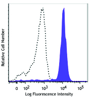
Human peripheral blood lymphocytes were stained with CD19 APC and HLA-DR (clone L243) PerCP/Cyanine5.5 (left), or mouse IgG2a, κ PerCP/Cyanine5.5 isotype control (right).
| Cat # | Size | Price | Quantity Check Availability | Save | ||
|---|---|---|---|---|---|---|
| 307629 | 25 tests | 141€ | ||||
| 307630 | 100 tests | 306€ | ||||
HLA-DR is a heterodimeric cell surface glycoprotein comprised of a 36 kD α (heavy) chain and a 27 kD β (light) chain. It is expressed on B cells, activated T cells, monocytes/macrophages, dendritic cells, and other non-professional APCs. In conjunction with the CD3/TCR complex and CD4 molecules, HLA-DR is critical for efficient peptide presentation to CD4+ T cells.
Product DetailsProduct Details
- Reactivity
- Human,Cynomolgus,Rhesus
- Antibody Type
- Monoclonal
- Host Species
- Mouse
- Formulation
- Phosphate-buffered solution, pH 7.2, containing 0.09% sodium azide and BSA (origin USA)
- Preparation
- The antibody was purified by affinity chromatography, and conjugated with PerCP/Cyanine5.5 under optimal conditions.
- Concentration
- Lot-specific (to obtain lot-specific concentration and expiration, please enter the lot number in our Certificate of Analysis online tool.)
- Storage & Handling
- The antibody solution should be stored undiluted between 2°C and 8°C, and protected from prolonged exposure to light. Do not freeze.
- Application
-
FC - Quality tested
- Recommended Usage
-
Each lot of this antibody is quality control tested by immunofluorescent staining with flow cytometric analysis. For flow cytometric staining, the suggested use of this reagent is 5 µl per million cells or 5 µl per 100 µl of whole blood. It is recommended that the reagent be titrated for optimal performance for each application.
* PerCP/Cyanine5.5 has a maximum absorption of 482 nm and a maximum emission of 690 nm. - Excitation Laser
-
Blue Laser (488 nm)
- Application Notes
-
The L243 monoclonal antibody reacts with the HLA-DR antigen, a member of MHC class II molecules. It does not cross react with HLA-DP and HLA-DQ. Clone L243 binds a conformational epitope on HLA-DRa which depends on the correct folding of the aß heterodimer.19
Additional reported applications (for the relevant formats) include: immunoprecipitation8, Western blotting8, in vitro blocking of mixed lymphocyte reactions9,10, depeletion of MHC class II cells7, immunohistochemical staining of acetone-fixed frozen sections4,5, and spatial biology (IBEX)21,22. For sensitive functional assays, we recommend using the Ultra-LEAF™ purified antibody (Endotoxin < 0.01 EU/µg, Azide-Free, 0.2 µm filtered) (Cat. No. 307648, 307665 - 307669). - Additional Product Notes
- BioLegend is in the process of converting the name PerCP/Cy5.5 to PerCP/Cyanine5.5. The dye molecule remains the same, so you should expect the same quality and performance from our PerCP/Cyanine5.5 products. Contact Technical Service if you have any questions.
-
Application References
(PubMed link indicates BioLegend citation) -
- Brodsky F. 1984. Immunogenetics 19:179.
- Robbins P, et al. 1987. Human Immunol. 18:301.
- Stites D, et al. 1986. Clin. Immunol. Immunopathol. 38:161.
- Warnke R, et al. 1980. J. Histochem. Cytochem. 28:771. (IHC)
- Engleman E, et al. 1981. P. Natl. Acad. Sci. USA 78:1791. (IHC)
- Zipf T, et al. 1981. Cancer Res. 41:4786.
- Goodier M, et al. 2000. J. Immunol. 165:139. (Depletion)
- Esser M, et al. 2001. J. Virol. 75:6173. (IP, WB)
- Kalka-Moll WM, et al. 2002. J. Immunol. 169:6149. (Block)
- Wang RF, et al. 1999. Science 284:1351. (Block)
- Zaba LC, et al. 2007. J. Exp. Med. 204:3183. PubMed
- Fujita H, et al. 2009. P. Natl. Acad. Sci. USA 106:21795. PubMed
- Charles N, et al. 2010. Nat. Med. 16:701. (FC) PubMed
- Goncalves RM, et al. 2010. Infect. Immun. 78:4763. PubMed
- Yoshino N, et al. 2000. Exp. Anim. (Tokyo) 49:97. (FC)
- Kim WK, et al. 2006. Am. J. Pathol. 168:822. (FC)
- Stein R, et al. 2011. Leuk. Lymphoma 52:273.
- Galkowska H, et al. 1996. Vet. Immunol. Immunopathol. 53:329.
- Moro M, et al. 2005. BMC Immunol. 6:24.
- Lauterbach N, et al. 2014. Mol Immunol. 59:19. PubMed
- Radtke AJ, et al. 2020. Proc Natl Acad Sci USA. 117:33455-33465. (SB) PubMed
- Radtke AJ, et al. 2022. Nat Protoc. 17:378-401. (SB) PubMed
- Product Citations
- RRID
-
AB_893575 (BioLegend Cat. No. 307629)
AB_893567 (BioLegend Cat. No. 307630)
Antigen Details
- Structure
- Ig superfamily, MHC class II, heterodimeric transmembrane protein, 36 kD heavy and 27 kD light chain
- Distribution
-
B cells, activated T cells, monocytes/macrophages, dendritic cells, other APCs
- Function
- Peptide presentation
- Ligand/Receptor
- CD3/TCR, CD4
- Cell Type
- Antigen-presenting cells, B cells, Dendritic cells, Macrophages, Monocytes, T cells, Tregs
- Biology Area
- Immunology, Innate Immunity
- Molecular Family
- MHC Antigens
- Antigen References
-
1. Levacher M, et al. 1990. Clin. Exp. Immunol. 81:177.
2. Terstappen L, et al. 1990. J. Leukocyte Biol. 48:138.
3. Edwards JA, et al. 1986. J. Immunol. 137:490.
4. van Es A, et al. 1984. Transplantation 37:65.
5. O'Doherty U, et al. 1994. Immunology 82:487.
6. Thomas R, et al. 1994. J. Immunol. 153:4016.
7. Grouard G, et al. 1996. Nature 384:364. - Gene ID
- 3122 View all products for this Gene ID 3123 View all products for this Gene ID
- UniProt
- View information about HLA-DR on UniProt.org
Related Pages & Pathways
Pathways


Related FAQs
- How stable is PerCP/Cyanine5.5 tandem as compared to PerCP alone?
-
PerCP/Cyanine5.5 is quite photostable and also better than PerCP alone in withstanding fixation.
Customers Also Purchased
Compare Data Across All Formats
This data display is provided for general comparisons between formats.
Your actual data may vary due to variations in samples, target cells, instruments and their settings, staining conditions, and other factors.
If you need assistance with selecting the best format contact our expert technical support team.


Follow Us