 Login / Register


- Clone
- BJ18 (See other available formats)
- Regulatory Status
- RUO
- Workshop
- VI A034
- Other Names
- Hermes, Pgp-1, H-CAM, HUTCH-1, ECMR III, gp85, Ly-24
- Isotype
- Mouse IgG1, κ
- Ave. Rating
- Submit a Review
- Product Citations
- publications

-

MDA-MB-231 cells were stained with anti-CD44 (clone BJ18), followed by Alexa Fluor® 546 secondary antibody and DAPI (nuclei). Images were aquired on a Nikon FC300 inverted microscope at 20X magnification. Data provided by Dr. John Nolan, La Jolla Bioengineering Institute.
| Cat # | Size | Price | Quantity Check Availability | Save | ||
|---|---|---|---|---|---|---|
| 338802 | 100 µg | 90€ | ||||
CD44 is a 80-95 kD glycoprotein also known as Hermes, Pgp1, H-CAM, or HUTCH. It is expressed on all leukocytes, endothelial cells, hepatocytes, and mesenchymal cells. As B and T cells become activated or progress to the memory stage, CD44 expression increases from a low or mid level of intensity to high expression levels. Thus, CD44 has been reported to be a valuable marker for memory cell subsets. CD44 is an adhesion molecule involved in leukocyte attachment to and rolling on endothelial cells, homing to peripheral lymphoid organs and to the sites of inflammation, and leukocyte aggregation.
Product DetailsProduct Details
- Reactivity
- Human
- Antibody Type
- Monoclonal
- Host Species
- Mouse
- Immunogen
- Normal human PBL
- Formulation
- Phosphate-buffered solution, pH 7.2, containing 0.09% sodium azide.
- Preparation
- The antibody was purified by affinity chromatography.
- Concentration
- 0.5 mg/ml
- Storage & Handling
- The antibody solution should be stored undiluted between 2°C and 8°C.
- Application
-
FC - Quality tested
CyTOF®, ICC - Verified - Recommended Usage
-
Each lot of this antibody is quality control tested by immunofluorescent staining with flow cytometric analysis. For flow cytometric staining, the suggested use of this reagent is ≤ 0.5 µg per million cells in 100 µl volume. It is recommended that the reagent be titrated for optimal performance for each application.
-
Application References
(PubMed link indicates BioLegend citation) -
- Kishimoto T, et al. eds. 1997 Leucocyte Typing VI:White Cell Differentiation Antigen. Garland Publishing Inc.
- Product Citations
- RRID
-
AB_1501199 (BioLegend Cat. No. 338802)
Antigen Details
- Structure
- Variable splicing of CD44 gene generates many CD44 isoforms, 85 kD
- Distribution
-
All leukocytes, epithelial cells, endothelial cells, hepatocytes, mesenchymal cells
- Function
- Leukocyte attachment and rolling on endothelial cells, stromal cells and ECM
- Ligand/Receptor
- Hyaluronan, MIP-1β, fibronectin, collagen
- Cell Type
- Endothelial cells, Epithelial cells, Leukocytes, Mesenchymal cells, Mesenchymal Stem Cells, Tregs
- Biology Area
- Cell Adhesion, Cell Biology, Immunology, Stem Cells
- Molecular Family
- Adhesion Molecules, CD Molecules
- Antigen References
-
1. Barclay AN, et al. 1997. The Leukocyte Antigen FactsBook Academic Press.
2. Haynes BF, et al. 1991. Cancer Cells 3:347.
3. Goldstein LA, et al. 1989. Cell 56:1063.
4. Mikecz K, et al. 1995. Nat. Med. 1:558. - Gene ID
- 960 View all products for this Gene ID
- UniProt
- View information about CD44 on UniProt.org


Customers Also Purchased
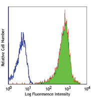
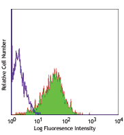
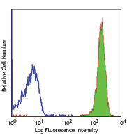
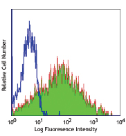
Compare Data Across All Formats
This data display is provided for general comparisons between formats.
Your actual data may vary due to variations in samples, target cells, instruments and their settings, staining conditions, and other factors.
If you need assistance with selecting the best format contact our expert technical support team.


Follow Us