 Login / Register


- Clone
- C8/144B (See other available formats)
- Regulatory Status
- RUO
- Other Names
- T8, Leu2
- Isotype
- Mouse IgG1, κ
- Ave. Rating
- Submit a Review
- Product Citations
- publications

-

Human paraffin-embedded tonsil tissue slices were prepared with a standard protocol of deparaffination and rehydration. Antigen retrieval was done with Tris-Buffered Saline 1X (1.0M, pH7.4) at 95°C for 40 minutes. Tissue was washed with PBS/ 0.05% Tween20 twice for five minutes and blocked with 5% FBS and 0.2% gelatin for 30 minutes. Then, the tissue was stained with 5 µg/ml of purified anti-human CD8a (clone C8/144B) at 4°C overnight. On the next day, the tissue was washed twice with PBS and stained with goat anti-mouse secondary antibody (clone Poly4053) DyLight™ 488 (green) for two hours at room temperature. The nuclei were counter staining with DAPI (blue). The image was captured with a 10X objective.
| Cat # | Size | Price | Quantity Check Availability | Save | ||
|---|---|---|---|---|---|---|
| 372902 | 100 µg | 48€ | ||||
CD8a is a 32-34 kD type I glycoprotein. It forms a homodimer (CD8a/a) or heterodimer (CD8a/b) with CD8b. CD8, also known as T8 and Leu2, is a member of the immunoglobulin superfamily found on the majority of thymocytes, a subset of peripheral blood T cells, and NK cells (which express almost exclusively CD8a homodimers). CD8 acts as a co-receptor with MHC class I-restricted T cell receptors in antigen recognition and T cell activation, and has been shown to play a role in thymic differentiation. Two domains in CD8a are important for function: the extracellular IgSF domain binds the α3 domain of MHC class I and the cytoplasmic CXCP motif binds the tyrosine kinase p56 Lck.
Product DetailsProduct Details
- Reactivity
- Human,Mouse,Rat
- Antibody Type
- Monoclonal
- Host Species
- Mouse
- Immunogen
- A 13 amino acid synthetic peptide from the C-terminal cytoplasmic domain of the alpha chain of the human CD8 molecule.
- Formulation
- Phosphate-buffered solution, pH 7.2, containing 0.09% sodium azide.
- Preparation
- The antibody was purified by affinity chromatography.
- Concentration
- 0.5 mg/ml
- Storage & Handling
- The antibody solution should be stored undiluted between 2°C and 8°C.
- Application
-
IHC-P - Quality tested
Activ, FC, IHC-F, WB - Reported in the literature, not verified in house - Recommended Usage
-
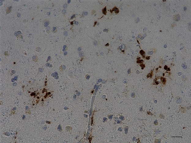
Each lot of this antibody is quality control tested by formalin-fixed paraffin-embedded immunohistochemical staining. For immunohistochemistry, a concentration range of 5.0 - 10 µg/ml is suggested. It is recommended that the reagent be titrated for optimal performance for each application.
- Application Notes
-
Additional reported applications (for the relevant formats) include activation3, flow cytometry4, immunohistochemical staining of frozen tissue sections2, and Western blotting5.
-
Application References
(PubMed link indicates BioLegend citation) -
- McQuitty E, et al. 2014. J. Cutan. Pathol. 41:88. (IHC-P)
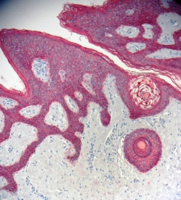
- Petukhova L, et al. 2010. Nature 466:113. (IHC-F)
- Clement M, et al. 2011. J. Immunol. 187:654. (Activ)
- MarTchal R, et al. 2010. BMC Cancer 10:340. (FC)
- Pontiggia L, et al. 2008. J. Invest. Dermatol. 129:480. (WB)
- Product Citations
- RRID
-
AB_2650657 (BioLegend Cat. No. 372902)
Antigen Details
- Structure
- Ig superfamily, homodimer or heterodimer with CD8β.
- Distribution
-
Majority of thymocytes, T cell subset, NK cells.
- Function
- MHC class I co-receptor, thymic differentiation, T-cell activation.
- Ligand/Receptor
- MHC class I molecules.
- Cell Type
- NK cells, T cells, Thymocytes
- Biology Area
- Immunology
- Molecular Family
- CD Molecules
- Antigen References
-
1. McQuitty E, et al. 2014. J. Cutan. Pathol. 41:88.
- Gene ID
- 100387674 View all products for this Gene ID
- UniProt
- View information about CD8a on UniProt.org


Customers Also Purchased
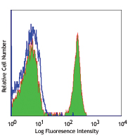
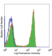
Compare Data Across All Formats
This data display is provided for general comparisons between formats.
Your actual data may vary due to variations in samples, target cells, instruments and their settings, staining conditions, and other factors.
If you need assistance with selecting the best format contact our expert technical support team.


Follow Us