 Login / Register


- Clone
- S17011E (See other available formats)
- Regulatory Status
- RUO
- Other Names
- CD16, CD32
- Isotype
- Rat IgG2b, κ
- Ave. Rating
- Submit a Review
- Product Citations
- publications

-

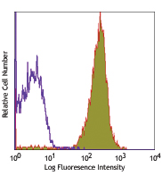
BALB/c splenocytes were incubated with TruStain FcX™ PLUS (clone S17011E, 0.25µg/106 cells, filled histogram) or TruStain FcX™ (clone 93, 1µg/106 cells, red line histogram) to block the Fc receptors, or were left untreated (black line histogram); then stained with CD90.2 (Thy-1.2, clone 53-2.1) FITC. Note the high background in the untreated cells and the bigger reduction in the background staining when the cells were incubated with TruStain FcX™ Plus compared to TruStain FcX™.
| Cat # | Size | Price | Quantity Check Availability | Save | ||
|---|---|---|---|---|---|---|
| 156603 | 50 µg | £45 | ||||
| 156604 | 500 µg | £189 | ||||
CD16 is the low affinity IgG Fc receptor III (FcR III) and CD32 is FcR II. CD16/CD32 are expressed on B cells, monocytes/macrophages, NK cells, granulocytes, mast cells, and dendritic cells. The Fc receptors bind antibody-antigen immune complexes and mediate adaptive immune responses. TruStain FcX™ PLUS is specific to the common epitope of CD16/CD32. It is useful for blocking non-specific binding of immunoglobulin to the Fc receptors and is more effective than TruStain FcX™.
Product DetailsProduct Details
- Verified Reactivity
- Mouse
- Antibody Type
- Monoclonal
- Host Species
- Rat
- Purity
- The antibody was purified by affinity chromatography.
- Formulation
- Phosphate-buffered solution, pH 7.2, containing 0.09% sodium azide.
- Concentration
- 0.5 mg/ml
- Storage & Handling
- The CD16/32 antibody solution should be stored undiluted between 2°C and 8°C.
- Application
-
FC - Quality tested
- Recommended Usage
-
For blocking of Fc receptors in flow cytometric analysis, pre-incubate the cells with TruStain FcX™ PLUS for 5-10 minutes, on ice, at 0.25 µg per 106 cells in a volume of 100 µl, prior to immunostaining. It is not necessary to wash the cells between the blocking and immunostaining steps.
- Application Notes
-
Clone S17011E blocks both clone 93 and 2.4G2 also raised against mouse CD16/32
- Product Citations
-
- RRID
-
AB_2783137 (BioLegend Cat. No. 156603)
AB_2783138 (BioLegend Cat. No. 156604)
Antigen Details
- Structure
- Ig superfamily, 40-60 kD
- Distribution
-
B cells, monocyte/macrophages, NK cells, neutrophils, mast cells, dendritic cells
- Ligand/Receptor
- IgG
- Cell Type
- B cells, Dendritic cells, Macrophages, Mast cells, Monocytes, Neutrophils, NK cells
- Biology Area
- Immunology
- Molecular Family
- CD Molecules, Fc Receptors
- Gene ID
- 14130 View all products for this Gene ID 14131 View all products for this Gene ID
- Specificity (DOES NOT SHOW ON TDS):
- CD16/32
- Specificity Alt (DOES NOT SHOW ON TDS):
- CD16/32
- App Abbreviation (DOES NOT SHOW ON TDS):
- FC
- UniProt
- View information about CD16/32 on UniProt.org


Other Formats
View All CD16/32 Reagents Request Custom Conjugation| Description | Clone | Applications |
|---|---|---|
| TruStain FcX™ PLUS (anti-mouse CD16/32) | S17011E | FC |
| APC/Cyanine7 anti-mouse CD16/32 | S17011E | FC |
| PE/Cyanine7 anti-mouse CD16/32 | S17011E | FC |
| PE anti-mouse CD16/32 | S17011E | FC |
| APC anti-mouse CD16/32 | S17011E | FC |
| FITC anti-mouse CD16/32 Antibody | S17011E | FC |
| PE/Dazzle™ 594 anti-mouse CD16/32 Antibody | S17011E | FC |
| PerCP/Cyanine5.5 anti-mouse CD16/32 Antibody | S17011E | FC |
| PE/Cyanine5 anti-mouse CD16/32 | S17011E | FC |
| Alexa Fluor® 700 anti-mouse CD16/32 | S17011E | FC |
| APC/Fire™ 750 anti-mouse CD16/32 | S17011E | FC |
| Brilliant Violet 510™ anti-mouse CD16/32 | S17011E | FC |
Customers Also Purchased
Compare Data Across All Formats
This data display is provided for general comparisons between formats.
Your actual data may vary due to variations in samples, target cells, instruments and their settings, staining conditions, and other factors.
If you need assistance with selecting the best format contact our expert technical support team.
-
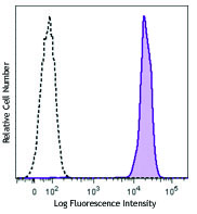
TruStain FcX™ PLUS (anti-mouse CD16/32)

BALB/c splenocytes were incubated with TruStain FcX™ PLUS (c... -
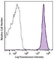
APC/Cyanine7 anti-mouse CD16/32

C57BL/6 mouse splenocytes stained with CD45R/B220 PE and CD1... -
PE/Cyanine7 anti-mouse CD16/32

C57BL/6 mouse splenocytes stained with CD45R/B220 APC and CD... -
PE anti-mouse CD16/32

C57BL/6 mouse splenocytes stained with CD45R/B220 APC and CD... -
APC anti-mouse CD16/32

C57BL/6 mouse splenocytes stained with CD45R/B220 PE and CD1... -
FITC anti-mouse CD16/32 Antibody

C57BL/6 mouse splenocytes were stained with anti-mouse CD45R... -
PE/Dazzle™ 594 anti-mouse CD16/32 Antibody

C57BL/6 mouse splenocytes were stained with anti-mouse CD45R... -
PerCP/Cyanine5.5 anti-mouse CD16/32 Antibody

C57BL/6 mouse splenocytes were stained with anti-mouse CD45R... -
PE/Cyanine5 anti-mouse CD16/32

C57BL/6 mouse splenocytes were stained with anti-mouse CD45R... -
Alexa Fluor® 700 anti-mouse CD16/32

C57BL/6 mouse splenocytes were stained with anti-mouse CD45R... -
APC/Fire™ 750 anti-mouse CD16/32

C57BL/6 mouse splenocytes were stained with anti-mouse CD45R... -
Brilliant Violet 510™ anti-mouse CD16/32

C57BL/6 splenocytes were stained with anti-mouse CD45R/B220 ...


Follow Us