 Login / Register


- Clone
- W16034A (See other available formats)
- Regulatory Status
- RUO
- Other Names
- Rab7, Ras-Related Protein Rab-7a, Ras-Associated Protein RAB7
- Isotype
- Rat IgG2a, κ
- Ave. Rating
- Submit a Review
- Product Citations
- publications

-

Western blot of anti-Rab7A antibody (clone W16034A) and isotype-matched IgG2A control. Blots were incubated with 2µg/ml (left) or IgG2A isotype control antibody (right) overnight at 4°C, followed by the incubation with horseradish peroxidase labeled goat anti-rat secondary antibody. Enhanced chemiluminescence was used as the detection system. M: Molecular weight marker; brain lysates: 20 µg; recombinant proteins: 10 ng. Purified anti-α-Tubulin (clone AA13) antibody was used as a loading control. Note that the band in the lane loaded with GST-Rab7A proteins in the blot probed with α-tubulin antibody (bottom left) is the remaining signal from the blot probed with anti-Rab7A antibody. Anti-GST antibody was used to confirm that GST-tagged recombinant proteins were loaded in the indicated lanes (data not shown). -

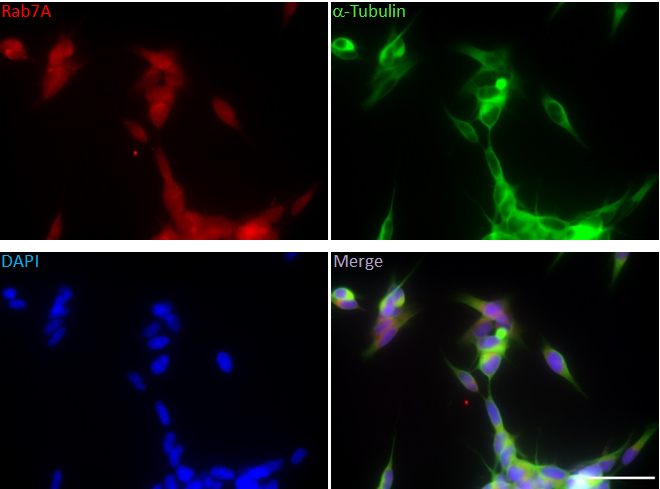
ICC staining of purified anti-Rab7A antibody (clone W16034A) on SH-SY5Y neuroblastoma cells. The cells were fixed with 4% PFA, permeabilized with 0.1% Triton X-100, and blocked with 2% normal goat serum and 0.02% BSA. The anti-Rab7A antibody (5 mg/ml) was pre-incubated with or without recombinant human Rab7A or Rab7B proteins (5 μg/ml) for 1 hour at room temperature prior to application to the cells for 24 hours at 4°C. The cells were co-stained with anti-α-Tubulin antibody (5 μg/ml; Cat. 909601; clone AA13), followed by incubation with Alexa Fluor® 594 anti-Rat (Rab7A, red) (Cat. 405422) and Alexa Fluor® 488 anti-mouse (α-Tubulin, green) (Cat. 405326) secondary antibodies for 1 hour at room temperature. Nuclei were counterstained with DAPI. Images were captured with a 40X objective. -

ICC staining of anti-Rab7A antibody (clone W16034A) on SH-SY5Y neuroblastoma cells. The cells were fixed with 4% PFA, permeabilized with 0.1% Triton X-100, and blocked with 2% normal goat serum and 0.02% BSA. The anti-Rab7A antibody (5 µg/ml) was pre-incubated with or without recombinant human Rab7A or Rab7B proteins (5 µg/ml) for one hour at room temperature prior to application to the cells for 24 hours at 4°C. The cells were co-stained with anti-α-Tubulin antibody (5 µg/ml; clone AA13), followed by incubation with Alexa Fluor® 594 anti-Rat (Rab7A, red) and Alexa Fluor® 488 anti-mouse (α-Tubulin, green) secondary antibodies for one hour at room temperature. Nuclei were counterstained with DAPI. Images were captured with a 40X objective. Scale bar: 50 µm. -

IHC staining of anti-Rab7A antibody (clone W16034A) on formalin-fixed paraffin-embedded normal human brain tissue. Following antigen retrieval using Sodium Citrate H.I.E.R, the tissue was incubated with the primary antibody at 5 µg/ml overnight at 4°C. Tissues were incubated with DAB for twenty minutes followed by hematoxylin counterstaining. Images were taken using 40X objectives. -

Direct ELISA of anti-Rab7A antibody (clone W16034A) binding to plate-immobilized recombinant human Rab7A, Rab7B, or PBS. ELISA was performed by coating wells with 150 ng of each recombinant protein for two hours at 37°C. Anti-Rab7A antibody was serially diluted and added to the plate with the concentration indicated on the x-axis, followed by incubation at 37°C for one hour. The plates were then incubated with horseradish peroxidase labeled goat anti-rat secondary antibody. TMB (3, 3', 5, 5' tetramethylbenzidine) was used as the detection system.
| Cat # | Size | Price | Quantity Check Availability | Save | ||
|---|---|---|---|---|---|---|
| 850401 | 25 µg | £85 | ||||
| 850402 | 100 µg | £213 | ||||
Rab7, a small GTPase of the Rab family, plays multiple roles including vesicular transport, endocytic trafficking, and autophagy. Rab7 is associated with both the endosome and lysosome, and it facilitates endosomal maturation, transport from the late endosome to the lysosome, and positioning of the endosome and lysosome via regulating their movement along cytoskeleton. Consistently, mutations or dysfunctions of Rab7 result in traffic disorders, which cause various diseases, such as neuropathy, cancer and lipid metabolism disease. Rab7 also plays important roles in microbial pathogen infection and survival, as well as in participating in the life cycle of viruses.
Product DetailsProduct Details
- Verified Reactivity
- Human, Rat, Mouse
- Antibody Type
- Monoclonal
- Host Species
- Rat
- Immunogen
- Full-length recombinant human Rab7A protein expressed in E. coli.
- Formulation
- Phosphate-buffered solution, pH 7.2, containing 0.09% sodium azide.
- Preparation
- The antibody was purified by affinity chromatography.
- Concentration
- 0.5 mg/ml
- Storage & Handling
- The antibody solution should be stored undiluted between 2°C and 8°C.
- Application
-
WB - Quality tested
Direct ELISA, ICC, IHC-P - Verified - Recommended Usage
-
Each lot of this antibody is quality control tested by Western blotting. For Western blotting, the suggested use of this reagent is 0.2 µg per ml. For immunocytochemistry, the suggested usage is 1.0 µg per ml. For immunohistochemical staining on formalin-fixed paraffin-embedded tissue sections, the suggested use of this reagent is 5.0 µg per ml. It is recommended that the reagent be titrated for optimal performance for each application.
- RRID
-
AB_2715874 (BioLegend Cat. No. 850401)
AB_2715874 (BioLegend Cat. No. 850402)
Antigen Details
- Structure
- Rab7A is a 207 amino acid protein with a molecular mass of 23 kD.
- Distribution
-
Tissue distribution: Rab7A is ubiquitously expressed.
Cellular distribution: Plasma membrane, cytosol, lysosome, endosome, and Golgi apparatus. - Function
- Rab7A is involved in the endocytic pathway and autophagy.
- Interaction
- Rab7A interacts with RILP, Rab1A, Rab11A, and UBC.
- Biology Area
- Cell Biology, Neurodegeneration, Neuroscience, Neuroscience Cell Markers, Protein Trafficking and Clearance
- Molecular Family
- Autophagosome Markers
- Antigen References
-
1. Guerra F, Bucci C. 2016. Cells. 5(3).
- Gene ID
- 7879 View all products for this Gene ID
- UniProt
- View information about Rab7A on UniProt.org

Other Formats
View All Rab7A Reagents Request Custom Conjugation| Description | Clone | Applications |
|---|---|---|
| Purified anti-Rab7A | W16034A | WB,Direct ELISA,ICC,IHC-P |
| Biotin anti-Rab7A | W16034A | WB |
| Alexa Fluor® 594 anti-Rab7A | W16034A | ICC |
Customers Also Purchased
Compare Data Across All Formats
This data display is provided for general comparisons between formats.
Your actual data may vary due to variations in samples, target cells, instruments and their settings, staining conditions, and other factors.
If you need assistance with selecting the best format contact our expert technical support team.
-
Purified anti-Rab7A

Western blot of anti-Rab7A antibody (clone W16034A) and isot... 
ICC staining of anti-Rab7A antibody (clone W16034A) on SH-SY... 
IHC staining of anti-Rab7A antibody (clone W16034A) on forma... 
Direct ELISA of anti-Rab7A antibody (clone W16034A) binding ... 
ICC staining of purified anti-Rab7A antibody (clone W16034A)... -
Biotin anti-Rab7A

Western blot of Biotin anti-Rab7A antibody (clone W16034A). ... -
Alexa Fluor® 594 anti-Rab7A

ICC staining of Alexa Fluor® 594 anti-Rab7A antibody (clone ...


Follow Us