 Login / Register


- Clone
- ICRF44 (See other available formats)
- Regulatory Status
- RUO
- Workshop
- IV M047
- Other Names
- Integrin αM chain, C3biR, CR3, Mac-1, Mo1, ITGAM
- Isotype
- Mouse IgG1, κ
- Ave. Rating
- Submit a Review
- Product Citations
- publications

-

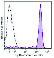
Human peripheral blood granulocytes were stained with CD11b (clone ICRF44) Brilliant Violet 421™ (filled histogram), or mouse IgG1, κ Brilliant Violet 421™ (open histogram). -

Human neutrophils were fixed with 1% paraformaldehyde (PFA) and then stained with 10 µg/ml CD11b (clone ICRF44) Brilliant Violet 421™ (red). Nuclei were counterstained with DRAQ5 (blue). The image was captured with a 40X objective
| Cat # | Size | Price | Quantity Check Availability | Save | ||
|---|---|---|---|---|---|---|
| 301323 | 25 tests | £140 | ||||
| 301324 | 100 tests | £261 | ||||
CD11b is a 165-170 kD type I transmembrane glycoprotein also known as αM integrin, Mac-1, CR3, and C3biR. CD11b non-covalently associates with integrin β2 (CD18) and is expressed on granulocytes, monocytes/macrophages, dendritic cells, NK cells, and subsets of T and B cells. CD11b/CD18 is critical for the transendothelial migration of monocytes and neutrophils. It is also involved in granulocyte adhesion, phagocytosis, and neutrophil activation. CD11b/CD18 interacts with ICAM-1 (CD54), ICAM-2 (CD102), ICAM-4, CD14, CD23, heparin, iC3b, fibrinogen, and factor X.
Product DetailsProduct Details
- Reactivity
- Human,Cynomolgus,Rhesus
- Antibody Type
- Monoclonal
- Host Species
- Mouse
- Formulation
- Phosphate-buffered solution, pH 7.2, containing 0.09% sodium azide and BSA (origin USA).
- Preparation
- The antibody was purified by affinity chromatography and conjugated with Brilliant Violet 421™ under optimal conditions.
- Concentration
- Lot-specific (to obtain lot-specific concentration and expiration, please enter the lot number in our Certificate of Analysis online tool.)
- Storage & Handling
- The antibody solution should be stored undiluted between 2°C and 8°C, and protected from prolonged exposure to light. Do not freeze.
- Application
-
FC - Quality tested
ICC - Verified - Recommended Usage
-
Each lot of this antibody is quality control tested by immunofluorescent staining with flow cytometric analysis. For flow cytometric staining, the suggested use of this reagent is 5 µL per million cells in 100 µL staining volume or 5 µL per 100 µL of whole blood.
Brilliant Violet 421™ excites at 405 nm and emits at 421 nm. The standard bandpass filter 450/50 nm is recommended for detection. Brilliant Violet 421™ is a trademark of Sirigen Group Ltd.
Learn more about Brilliant Violet™.
This product is subject to proprietary rights of Sirigen Inc. and is made and sold under license from Sirigen Inc. The purchase of this product conveys to the buyer a non-transferable right to use the purchased product for research purposes only. This product may not be resold or incorporated in any manner into another product for resale. Any use for therapeutics or diagnostics is strictly prohibited. This product is covered by U.S. Patent(s), pending patent applications and foreign equivalents. - Excitation Laser
-
Violet Laser (405 nm)
- Application Notes
-
The ICRF44 antibody inhibits heterotypic adhesion of granulocytes in response to fMLP. Additional reported applications (for the relevant formats) include: immunohistochemical staining of acetone-fixed frozen tissue sections, immunofluorescence microscopy5, stimulation of monocytes3, blocking of heterotypic PMN aggregation8, and blocking of granulocyte activation12. This clone was tested in-house and does not work on formalin fixed paraffin-embedded (FFPE) tissue.
The Ultra-LEAF™ purified antibody (Endotoxin < 0.01 EU/µg, Azide-Free, 0.2 µm filtered) is recommended for functional assays (Cat. Nos. 301361 & 301362). -
Application References
(PubMed link indicates BioLegend citation) -
- Knapp W. 1989. Leucocyte Typing IV. Oxford University Press New York.
- Barclay N, et al. 1997. The Leucocyte Antigen Facts Book. Academic Press Inc. San Diego.
- Rezzonico R, et al. 2001. Blood 97:2932. (Stim)
- Marsik C, et al. 2003. Shock 20:493. (FC)
- David A, et al. 2003. J. Leukoc. Biol. 74:551. (IF)
- Charles N, et al. 2010. Nat. Med. 16:701. (FC) PubMed
- Thurlow LR, et al. 2010. Infect. Immun. 128:1128. (FC) PubMed
- Jadhav S, et al. 2001. J. Immunol. 167:5986. (Block)
- Yoshino N, et al. 2000. Exp. Anim. (Tokyo) 49:97. (FC)
- Sestak K, et al. 2007. Vet. Immunol. Immunopathol. 119:21. (FC)
- Wen T, et al. 2014. J Immunol. 192:5481. (FC) PubMed
- Sprong T, et al. 2003. Blood 102:3702. (Block)
- Cash JL, et al. 2013. EMBO Rep. 14:999. (FC) PubMed
- Larsson K, et al. 2015. PNAS. PubMed
- Product Citations
- RRID
-
AB_10933087 (BioLegend Cat. No. 301323)
AB_11219589 (BioLegend Cat. No. 301324)
Antigen Details
- Structure
- Integrin, type I transmembrane glycoprotein, associates with integrin β2 (CD18), 165-170 kD
- Distribution
-
Granulocytes, monocytes/macrophages, dendritic cells, NK cells, subset of T cells, subset of B cells
- Function
- Adhesion, phagocytosis, chemotaxis, neutrophil activation
- Ligand/Receptor
- ICAM-1(CD54), ICAM-2 (CD102), ICAM-4, CD14, CD23, heparin, iC3b, fibrinogen, factor X
- Cell Type
- B cells, Dendritic cells, Granulocytes, Macrophages, Monocytes, Neutrophils, NK cells, T cells, Tregs
- Biology Area
- Cell Adhesion, Cell Biology, Costimulatory Molecules, Immunology, Innate Immunity, Neuroscience, Neuroscience Cell Markers
- Molecular Family
- Adhesion Molecules, CD Molecules
- Antigen References
-
1. Stewart M, et al. 1995. Curr Opin Cell Biol. 7:690.
- Gene ID
- 3684 View all products for this Gene ID
- UniProt
- View information about CD11b on UniProt.org
Related Pages & Pathways
Pathways


Related FAQs
- What is the F/P ratio range of our BV421™ format antibody reagents?
-
It is lot-specific. On average it ranges between 2-4.
Customers Also Purchased
Compare Data Across All Formats
This data display is provided for general comparisons between formats.
Your actual data may vary due to variations in samples, target cells, instruments and their settings, staining conditions, and other factors.
If you need assistance with selecting the best format contact our expert technical support team.


Follow Us