 Login / Register


- Clone
- 16-10A1 (See other available formats)
- Regulatory Status
- RUO
- Other Names
- B7-1, B7, Ly-53
- Isotype
- Armenian Hamster IgG
- Ave. Rating
- Submit a Review
- Product Citations
- publications

-

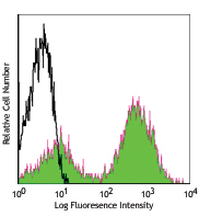
LPS-stimulated (day 3) C57BL/6 mouse splenocytes stained with CD80 (clone 16-10A1) Brilliant Violet 421™ (filled histogram) or Armenian hamster IgG Brilliant Violet 421™ isotype control (open histogram). -

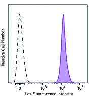
C57BL/6 mouse frozen thymus section was fixed with 4% paraformaldehyde (PFA) for 10 minutes at room temperature and blocked with 5% FBS for 30 minutes at room temperature. Then the section was stained with 10 µg/ml of CD80 (clone 16-10A1) Brilliant Violet® 421 (purple), 10 µg/ml of CD90.2 (clone 30-H12) Alexa Fluor® 647 (red) and 10 µg/ml of CD326 (clone G8.8) Alexa Fluor® 594 (green) overnight at 4°C. The image was captured by 10X objective.
| Cat # | Size | Price | Quantity Check Availability | Save | ||
|---|---|---|---|---|---|---|
| 104725 | 125 µL | £140 | ||||
| 104726 | 500 µL | £275 | ||||
CD80 is a 60 kD highly glycosylated protein. It is a member of the Ig superfamily and is also known as B7-1, B7, and Ly-53. CD80 is constitutively expressed on dendritic cells and monocytes/macrophages, and inducibly expressed on activated B and T cells. The ligation of CD28 on T cells with CD80 and CD86 (B7-2) on antigen presenting cells (such as dendritic cells, macrophages, and B cells) elicits co-stimulation of T cells resulting in enhanced cell activation, proliferation, and cytokine production. CD80 appears to be expressed later in the immune response than CD86. CD80 can also bind to CD152, also known as CTLA-4, to deliver an inhibitory signal to T cells.
Product DetailsProduct Details
- Reactivity
- Mouse
- Antibody Type
- Monoclonal
- Host Species
- Armenian Hamster
- Immunogen
- CHO cell line transfected with mouse B7 (CD80)
- Formulation
- Phosphate-buffered solution, pH 7.2, containing 0.09% sodium azide and BSA (origin USA).
- Preparation
- The antibody was purified by affinity chromatography and conjugated with Brilliant Violet 421™ under optimal conditions.
- Concentration
- Lot-specific (to obtain lot-specific concentration and expiration, please enter the lot number in our Certificate of Analysis online tool.)
- Storage & Handling
- The antibody solution should be stored undiluted between 2°C and 8°C, and protected from prolonged exposure to light. Do not freeze.
- Application
-
FC - Quality tested
IHC-F - Verified - Recommended Usage
-
Each lot of this antibody is quality control tested by immunofluorescent staining with flow cytometric analysis. For flow cytometric staining, the suggested use of this reagent is 5 µl per million cells in 100 µl staining volume or 5 µl per 100 µl of whole blood. For immunohistochemical staining on frozen tissue sections, the suggested use is 5-10 µg/mL. It is recommended that the reagent be titrated for optimal performance for each application.
Brilliant Violet 421™ excites at 405 nm and emits at 421 nm. The standard bandpass filter 450/50 nm is recommended for detection. Brilliant Violet 421™ is a trademark of Sirigen Group Ltd.
Learn more about Brilliant Violet™.
This product is subject to proprietary rights of Sirigen Inc. and is made and sold under license from Sirigen Inc. The purchase of this product conveys to the buyer a non-transferable right to use the purchased product for research purposes only. This product may not be resold or incorporated in any manner into another product for resale. Any use for therapeutics or diagnostics is strictly prohibited. This product is covered by U.S. Patent(s), pending patent applications and foreign equivalents. - Excitation Laser
-
Violet Laser (405 nm)
- Application Notes
-
Additional reported applications (for the relevant formats) include: immunoprecipitation2, in vitro and in vivo blocking of CTLA-4 Ig to CD80 by blocking costimulation of T cells by activated B cells2-4, and immunohistochemical staining of acetone-fixed frozen sections1,4. The Ultra-LEAF™ purified antibody (Endotoxin < 0.01 EU/µg, Azide-Free, 0.2 µm filtered) is recommended for functional assays (Cat. Nos. 104747-104752).
-
Application References
(PubMed link indicates BioLegend citation) -
- Harlan DM, et al. 1994. P. Natl. Acad. Sci. USA 91:3137. (IHC)
- Razi-Wolf Z, et al. 1992. P. Natl. Acad. Sci. USA 89:4210. (Block, IP)
- Hathcock KS, et al. 1994. J. Exp. Med. 180:631. (Block)
- Herold KC, et al. 1997. J. Immunol. 158:984. (Block, IHC)
- Ma XT, et al. 2006. Cancer Res. 66:1169.
- Andoniou CE, et al. 2005. Nature Immunology 6:1011. (FC)
- Lawson BR, et al. 2007. J. Immunol. 178:5366.
- Turnquist HR, et al. 2007. J. Immunol. 178:7018.
- Misra RS, et al. 2010. J. Exp Med. 207:1775. PubMed
- del Rio ML, et al. 2011. Transpl. Int. 24:501. (FC) PubMed
- Philipsen L, et al. 2013. Mol Cell Proteomics. 12:2551. PubMed
- Product Citations
- RRID
-
AB_10900989 (BioLegend Cat. No. 104725)
AB_2561445 (BioLegend Cat. No. 104726)
Antigen Details
- Structure
- Ig superfamily, 60 kD
- Distribution
-
Macrophages, activated B cells and T cells, dendritic cells
- Function
- T cell costimulation
- Ligand/Receptor
- CD28 (stimulatory), CD152(CTLA4) (inhibitory)
- Cell Type
- B cells, Dendritic cells, Macrophages, T cells, Tregs
- Biology Area
- Cell Biology, Costimulatory Molecules, Immunology, Neuroscience, Neuroscience Cell Markers
- Molecular Family
- CD Molecules, Immune Checkpoint Receptors
- Antigen References
-
1. Barclay AN, et al. 1997. The Leukocyte Antigen FactsBook Academic Press.
2. Linsley PS, et al. 1991. J. Exp. Med. 174:561.
3. Salomon B, et al. 2001. Annu. Rev. Immunol. 19:225. - Gene ID
- 12519 View all products for this Gene ID
- UniProt
- View information about CD80 on UniProt.org
Related Pages & Pathways
Pathways


Related FAQs
- What is the F/P ratio range of our BV421™ format antibody reagents?
-
It is lot-specific. On average it ranges between 2-4.
Customers Also Purchased
Compare Data Across All Formats
This data display is provided for general comparisons between formats.
Your actual data may vary due to variations in samples, target cells, instruments and their settings, staining conditions, and other factors.
If you need assistance with selecting the best format contact our expert technical support team.


Follow Us