FITC anti-human CD14 Antibody

Pricing & Availability
Clone
HCD14 (See other available formats)
Regulatory Status
RUO
Other Names
LPS receptor
Isotype
Mouse IgG1, κ
Ave. Rating
Submit a Review
Product Citations
publications
HCD14_FITC_061407
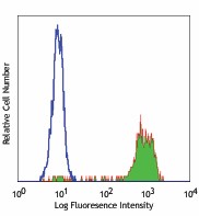
Human peripheral blood monocytes stained with HCD14 FITC
  • HCD14_FITC_061407
    Human peripheral blood monocytes stained with HCD14 FITC
See FITC spectral data
Cat # Size Price Quantity Check Availability Save
325603 25 tests £65
Check Availability


Need larger quantities of this item?
Request Bulk Quote
325604 100 tests £145
Check Availability


Need larger quantities of this item?
Request Bulk Quote
Description

CD14 is a 53-55 kD glycosylphosphatidylinositol (GPI)-linked membrane glycoprotein also known as LPS receptor. CD14 is expressed at high levels on monocytes and macrophages, and at lower levels on granulocytes. Some dendritic cell populations such as interfollicular dendritic cells, reticular dendritic cells, and Langerhans cells have also been reported to express CD14. As a high-affinity receptor for LPS, CD14 is involved in the clearance of gram-negative pathogens and in the upregulation of adhesion molecules and cytokines expression in monocytes and neutrophils.

Product Details
Technical Data Sheet (pdf)

Product Details

Reactivity
Human
Antibody Type
Monoclonal
Host Species
Mouse
Formulation
Phosphate-buffered solution, pH 7.2, containing 0.09% sodium azide and BSA (origin USA)
Preparation
The antibody was purified by affinity chromatography, and conjugated with FITC under optimal conditions.
Concentration
Lot-specific (to obtain lot-specific concentration and expiration, please enter the lot number in our Certificate of Analysis online tool.)
Storage & Handling
The antibody solution should be stored undiluted between 2°C and 8°C, and protected from prolonged exposure to light. Do not freeze.
Application

FC - Quality tested

Recommended Usage

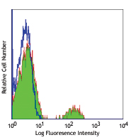
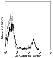
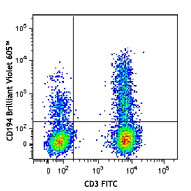
Each lot of this antibody is quality control tested by immunofluorescent staining with flow cytometric analysis. For flow cytometric staining, the suggested use of this reagent is 5 µl per million cells in 100 µl staining volume or 5 µl per 100 µl of whole blood.

Excitation Laser
Blue Laser (488 nm)
Application Notes

Additional reported applications (for the relevant formats) include: immunofluorescence microscopy. This clone was tested in-house and does not work on formalin fixed paraffin-embedded (FFPE) tissue.

Application References

(PubMed link indicates BioLegend citation)
  1. McMichael A, et al. 1987. Leucocyte Typing III. Oxford University Press. New York.
  2. Knapp W, et al. Eds. 1989. Leucocyte Typing IV. Oxford University Press. New York.
  3. Schlossman S, et al. Eds. 1995. Leucocyte Typing V. Oxford University Press. New York.
Product Citations
  1. BÖiers C, et al. 2018. Dev Cell. 44:362. PubMed
  2. Setliff I, et al. 2018. Cell Host Microbe. 23:845. PubMed
  3. Hansen IS, et al. 2018. Nat Commun. 9:863. PubMed
  4. Toepfner N et al. 2018. eLife. 7 pii: e29213. PubMed
  5. Alissafi T, et al. 2018. J Clin Invest. 128:3840. PubMed
  6. Walk J, et al. 2019. Nat Commun. 10:874. PubMed
  7. Yang H, et al. 2019. Mol Metab. 23:24. PubMed
  8. Sajadi MM, et al. 2018. Cell. 173:1783. PubMed
  9. Deniset JF et al. 2019. Immunity. 51(1):131-140 . PubMed
  10. Sykes DB et al. 2016. Cell. 167(1):171-186 . PubMed
  11. Beyer AI, et al. 2017. Stem Cells Dev. 26:102. PubMed
  12. Horn LA, et al. 2017. Oncotarget. 8:57964. PubMed
  13. Martin GE, et al. 2018. Front Immunol. 1.019444444. PubMed
  14. Hazenberg MD, et al. 2019. Blood Adv. 2.659722222. PubMed
  15. Domingos-Pereira S, et al. 2019. Cancer Immunol Res. 7:621. PubMed
  16. Matsuyama H, et al. 2019. Sci Rep. 9:13181. PubMed
  17. Bennstein SB, et al. 2020. eLife. 9:e55232.. PubMed
  18. Timmerman K, et al. 2008. J Leukoc Biol. 84:1271. PubMed
  19. Huang Y, et al. 2008. J Immunol. 180:2367. PubMed
  20. Mangsbo S, et al. 2009. J Immunol. 183:6724. PubMed
  21. Jensen M, et al. 2010. Mult Scler. 16:30. PubMed
  22. Švajger U, et al. 2009. Immunology. 129:525. PubMed
  23. Zhu H, et al. 2014. Lupus. 23:624. PubMed
  24. Lepore M, et al. 2014. J Exp Med. 211:1363. PubMed
  25. Mandl M, et al. 2014. PLoS One. 9:112140. PubMed
  26. Bettman N, et al. 2015. Blood Cells Mol Dis. 55: 180-186. PubMed
  27. Jama B, Morris G 2014. J Vis Exp. 93: 52257. PubMed
  28. Bal S, et al. 2016. Nat Immunol. 10.1038/ni.3444. PubMed
  29. An L, et al. 2016. Sci Rep. 6: 33346. PubMed
  30. Jimenez-Duran G, et al. 2020. EBioMedicine. 61:103039. PubMed
  31. Tan EE, et al. 2020. J Clin Invest. 130:5817. PubMed
  32. Ramaseri Sunder S, et al. 2020. J Interferon Cytokine Res. 40:310. PubMed
  33. Cizmeci D, et al. 2021. eLife. 10:00. PubMed
  34. Esaulova E, et al. 2020. Cell Host Microbe. 29(2):165-178.e8. PubMed
  35. Shen X, et al. 2020. Molecular Cell. 80(6):1013-1024.e6. PubMed
  36. Wang F, et al. 2021. Cell. 184(2):422-440.e17. PubMed
  37. Golebski K, et al. 2021. Immunity. 54(2):291-307.e7. PubMed
  38. Rossignol ED, et al. 2021. Cell Reports. 35(8):109167. PubMed
  39. Kerr SC, et al. 2020. Clin Exp Allergy. 50:904. PubMed
  40. Pholcharee T, et al. 2021. Nat Commun. 12:1063. PubMed
  41. Wißfeld J, et al. 2021. Glia. 69:1393. PubMed
  42. Laustsen A, et al. 2021. Elife. 10:. PubMed
  43. Georgakis S, et al. 2021. JCI Insight. 6:. PubMed
  44. Lu J, et al. 2021. Front Immunol. 12:732612. PubMed
  45. Witkowski M, et al. 2021. Nature. 600:295. PubMed
  46. Aram J, et al. 2020. Biomedicines. 8:. PubMed
  47. Chen IP, et al. 2020. Bio Protoc. 10:e3854. PubMed
  48. Kokkinopoulos I, et al. 2021. Sci Rep. 11:15759. PubMed
  49. Kato M, et al. 2021. PLoS One. 16:e0252116. PubMed
  50. Warmuth S, et al. 2022. Oncoimmunology. 10:2004661. PubMed
  51. Feng T, et al. 2022. Nat Commun. 13:5208. PubMed
  52. Maas RR, et al. 2021. Nat Protoc. 16:4692. PubMed
  53. Bradley D, et al. 2022. Nat Commun. 13:5606. PubMed
  54. Huang H, et al. 2022. Nat Commun. 13:5925. PubMed
  55. Zhang P, et al. 2022. BMC Med. 20:435. PubMed
  56. Bhattacharya S, et al. 2022. J Clin Invest. Online ahead of print. PubMed
  57. Raggi C, et al. 2022. Curr Protoc. 2:e389. PubMed
  58. Asashima H, et al. 2022. J Clin Invest. 132: . PubMed
  59. Lin L, et al. 2023. Mol Ther Nucleic Acids. 31:88. PubMed
  60. Jiang Y, et al. 2023. Sci Adv. 9:eadc8933. PubMed
  61. Seegren PV, et al. 2023. Nat Aging. . PubMed
RRID
AB_830676 (BioLegend Cat. No. 325603)
AB_830677 (BioLegend Cat. No. 325604)

Antigen Details

Structure
GPI-linked membrane glycoprotein, 53-55 kD
Distribution

Monocytes, macrophages, granulocytes (low)

Function
LPS receptor, clearance of Gram-negative pathogens
Ligand/Receptor
LPS
Cell Type
Granulocytes, Macrophages, Monocytes, Neutrophils
Biology Area
Cell Biology, Immunology, Innate Immunity, Neuroinflammation, Neuroscience
Molecular Family
CD Molecules
Antigen References

1. Stocks S, et al. 1990. Biochem. J. 268:275.
2. Wright S, et al. 1990. Science 249:1434.

Gene ID
929 View all products for this Gene ID
UniProt
View information about CD14 on UniProt.org

Related FAQs

There are no FAQs for this product.
Go To Top Version: 2    Revision Date: 07/13/2015

8999 BioLegend Way, San Diego, CA 92121 www.biolegend.com
Toll-Free Phone: 1-877-Bio-Legend (246-5343) Phone: (858) 768-5800 Fax: (877) 455-9587

This data display is provided for general comparisons between formats.
Your actual data may vary due to variations in samples, target cells, instruments and their settings, staining conditions, and other factors.
If you need assistance with selecting the best format contact our expert technical support team.

ProductsHere

Login / Register
Remember me
Forgot your password? Reset password?
Create an Account