 Login / Register


- Clone
- RPA-T4 (See other available formats)
- Regulatory Status
- RUO
- Workshop
- IV T114
- Other Names
- T4
- Isotype
- Mouse IgG1, κ
- Ave. Rating
- Submit a Review
- Product Citations
- publications

-

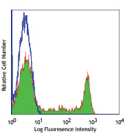
Human peripheral blood lymphocytes were stained with CD4 (clone RPA-T4) Brilliant Violet 510™ (filled histogram) or mouse IgG1, κ Brilliant Violet 510™ isotype control (open histogram).
| Cat # | Size | Price | Quantity Check Availability | Save | ||
|---|---|---|---|---|---|---|
| 300545 | 25 tests | 150€ | ||||
| 300546 | 100 tests | 340€ | ||||
CD4, also known as T4, is a 55 kD single-chain type I transmembrane glycoprotein expressed on most thymocytes, a subset of T cells, and monocytes/macrophages. CD4, a member of the Ig superfamily, recognizes antigens associated with MHC class II molecules, and participates in cell-cell interactions, thymic differentiation, and signal transduction. CD4 acts as a primary receptor for HIV, binding to HIV gp120. CD4 has also been shown to interact with IL-16.
Product DetailsProduct Details
- Reactivity
- Human
- Antibody Type
- Monoclonal
- Host Species
- Mouse
- Formulation
- Phosphate-buffered solution, pH 7.2, containing 0.09% sodium azide and BSA (origin USA).
- Preparation
- The antibody was purified by affinity chromatography and conjugated with Brilliant Violet 510™ under optimal conditions.
- Concentration
- Lot-specific (to obtain lot-specific concentration and expiration, please enter the lot number in our Certificate of Analysis online tool.)
- Storage & Handling
- The antibody solution should be stored undiluted between 2°C and 8°C, and protected from prolonged exposure to light. Do not freeze.
- Application
-
FC - Quality tested
- Recommended Usage
-
Each lot of this antibody is quality control tested by immunofluorescent staining with flow cytometric analysis. For flow cytometric staining, the suggested use of this reagent is 5 µl per million cells in 100 µl staining volume or 5 µl per 100 µl of whole blood.
Brilliant Violet 510™ excites at 405 nm and emits at 510 nm. The bandpass filter 510/50 nm is recommended for detection, although filter optimization may be required depending on other fluorophores used. Be sure to verify that your cytometer configuration and software setup are appropriate for detecting this channel. Refer to your instrument manual or manufacturer for support. Brilliant Violet 510™ is a trademark of Sirigen Group Ltd.
Learn more about Brilliant Violet™.
This product is subject to proprietary rights of Sirigen Inc. and is made and sold under license from Sirigen Inc. The purchase of this product conveys to the buyer a non-transferable right to use the purchased product for research purposes only. This product may not be resold or incorporated in any manner into another product for resale. Any use for therapeutics or diagnostics is strictly prohibited. This product is covered by U.S. Patent(s), pending patent applications and foreign equivalents. - Excitation Laser
-
Violet Laser (405 nm)
- Application Notes
-
The RPA-T4 antibody binds to the D1 domain of CD4 (CDR1 and CDR3 epitopes) and can block HIV gp120 binding and inhibit syncytia formation. Additional reported applications (for the relevant formats) include: immunohistochemistry of acetone-fixed frozen sections3,4,5, blocking of T cell activation1,2, and spatial biology (IBEX)10,11. This clone was tested in-house and does not work on formalin fixed paraffin-embedded (FFPE) tissue. The Ultra-LEAF™ purified antibody (Endotoxin < 0.01 EU/µg, Azide-Free, 0.2 µm filtered) is recommended for functional assays (Cat. No. 300569 - 300574).
- Application References
-
- Knapp W, et al. 1989. Leucocyte Typing IV. Oxford University Press. New York. (Activ)
- Moir S, et al. 1999. J. Virol. 73:7972. (Activ)
- Deng MC, et al. 1995. Circulation 91:1647. (IHC)
- Friedman T, et al. 1999. J. Immunol. 162:5256. (IHC)
- Mack CL, et al. 2004. Pediatr. Res. 56:79. (IHC)
- Lan RY, et al. 2006. Hepatology 43:729.
- Zenaro E, et al. 2009. J. Leukoc. Biol. 86:1393. (FC) PubMed
- Yoshino N, et al. 2000. Exp. Anim. (Tokyo) 49:97. (FC)
- Stoeckius M, et al. 2017. Nat. Methods. 14:865. (PG)
- Radtke AJ, et al. 2020. Proc Natl Acad Sci USA. 117:33455-33465. (SB) PubMed
- Radtke AJ, et al. 2022. Nat Protoc. 17:378-401. (SB) PubMed
- Product Citations
- RRID
-
AB_2563313 (BioLegend Cat. No. 300545)
AB_2563314 (BioLegend Cat. No. 300546)
Antigen Details
- Structure
- Ig superfamily, type I transmembrane glycoprotein, 55 kD
- Distribution
-
T cell subset, majority of thymocytes, monocytes/macrophages
- Function
- MHC class II co-receptor, lymphocyte adhesion, thymic differentiation, HIV receptor
- Ligand/Receptor
- MHC class II molecules, HIV gp120, IL-16
- Cell Type
- Dendritic cells, Macrophages, Monocytes, T cells, Thymocytes, Tregs
- Biology Area
- Immunology
- Molecular Family
- CD Molecules
- Antigen References
-
1. Center D, et al. 1996. Immunol. Today 17:476.
2. Gaubin M, et al. 1996. Eur. J. Clin. Chem. Clin. Biochem. 34:723. - Gene ID
- 920 View all products for this Gene ID
- UniProt
- View information about CD4 on UniProt.org
Related Pages & Pathways
Pathways


Related FAQs
- I am unable to see expression of T cell markers such as CD3 and CD4 post activation.
- TCR-CD3 complexes on the T-lymphocyte surface are rapidly downregulated upon activation with peptide-MHC complex, superantigen or cross-linking with anti-TCR or anti-CD3 antibodies. PMA/Ionomycin treatment has been shown to downregulate surface CD4 expression. Receptor downregulation is a common biological phenomenon and so make sure that your stimulation treatment is not causing it in your sample type.
Customers Also Purchased
Compare Data Across All Formats
This data display is provided for general comparisons between formats.
Your actual data may vary due to variations in samples, target cells, instruments and their settings, staining conditions, and other factors.
If you need assistance with selecting the best format contact our expert technical support team.


Follow Us