 Login / Register


- Clone
- 15-2 (See other available formats)
- Regulatory Status
- RUO
- Other Names
- MMR (macrophage mannose receptor), MR (mannose receptor), CD206, MRC1
- Isotype
- Mouse IgG1, κ
- Ave. Rating
- Submit a Review
- Product Citations
- publications
| Cat # | Size | Price | Quantity Check Availability | Save | ||
|---|---|---|---|---|---|---|
| 321141 | 25 tests | 172€ | ||||
| 321142 | 100 tests | 335€ | ||||
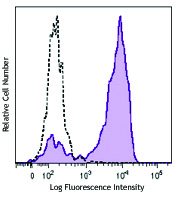
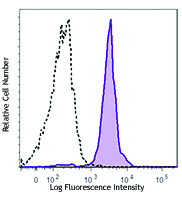
Macrophage mannose receptor (MMR) is a 162-175 kD type I membrane protein also known as CD206, MRC1, or mannose receptor (MR). It is a pattern recognition receptor (PRR) that belongs to C-type lectin superfamily. MMR is expressed on macrophages, dendritic cells, and hepatic or lymphatic endothelial cells, but not on monocytes. MMR recognizes a range of microbial carbohydrates bearing mannose, fucose, or N-acetyl glucosamine. MMR mediates endocytosis and phagocytosis, induces activation of macrophages and antigen presentation, plays an important role in host defense, and provides a link between innate and adaptive immunity.
Product DetailsProduct Details
- Reactivity
- Human
- Antibody Type
- Monoclonal
- Host Species
- Mouse
- Immunogen
- Purified human mannose receptor
- Formulation
- Phosphate-buffered solution, pH 7.2, containing 0.09% sodium azide and BSA (origin USA).
- Preparation
- The antibody was purified by affinity chromatography and conjugated with Brilliant Violet 785™ under optimal conditions.
- Concentration
- Lot-specific (to obtain lot-specific concentration and expiration, please enter the lot number in our Certificate of Analysis online tool.)
- Storage & Handling
- The antibody solution should be stored undiluted between 2°C and 8°C, and protected from prolonged exposure to light. Do not freeze.
- Application
-
FC - Quality tested
- Recommended Usage
-
Each lot of this antibody is quality control tested by immunofluorescent staining with flow cytometric analysis. For flow cytometric staining, the suggested use of this reagent is 5 µl per million cells in 100 µl staining volume or 5 µl per 100 µl of whole blood.
Brilliant Violet 785™ excites at 405 nm and emits at 785 nm. The bandpass filter 780/60 nm is recommended for detection, although filter optimization may be required depending on other fluorophores used. Be sure to verify that your cytometer configuration and software setup are appropriate for detecting this channel. Refer to your instrument manual or manufacturer for support. Brilliant Violet 785™ is a trademark of Sirigen Group Ltd.
Learn more about Brilliant Violet™.
This product is subject to proprietary rights of Sirigen Inc. and is made and sold under license from Sirigen Inc. The purchase of this product conveys to the buyer a non-transferable right to use the purchased product for research purposes only. This product may not be resold or incorporated in any manner into another product for resale. Any use for therapeutics or diagnostics is strictly prohibited. This product is covered by U.S. Patent(s), pending patent applications and foreign equivalents. - Excitation Laser
-
Violet Laser (405 nm)
- Application Notes
-
The 15-2 antibody blocks the interaction of MMR with its ligand, and inhibits mannose receptor-mediated degradation of t-PA by macrophages. Additional reported applications of this antibody (for the relevant formats) include: Western blotting1, blocking of ligand binding1,2, immunofluorescence3, and immunohistochemical staining of acetone-fixed frozen tissue sections1. The Utra-LEAF™ purified antibody (Endotoxin < 0.01 EU/µg, Azide-Free, 0.2 µm filtered) is recommended for functional assays (Cat. No. 321149 and 321150).
- Application References
-
- Noorman F, et al. 1997. J. Leukocyte Biol. 61:63. (WB, IHC, Block)
- Barrett-Bergshoeff M, et al. 1997. Thromb Haemost. 77:718. (Block)
- Kato M, et al. 2007. J. Immunol. 179:6052. (IF)
- Product Citations
- RRID
-
AB_2734301 (BioLegend Cat. No. 321141)
AB_2734302 (BioLegend Cat. No. 321142)
Antigen Details
- Structure
- Type I membrane protein, Pattern Recognition Receptor (PRR) family, C-type lectin superfamily, 162-175 kD
- Distribution
-
Macrophages, dendritic cells, hepatic and lymphatic endothelial cells
- Function
- Pathogen binding, facilitate phagocytosis and endocytosis, macrophage activation and antigen presentation
- Ligand/Receptor
- Mannose, fucose, N-acetyl glucosamine
- Cell Type
- Dendritic cells, Endothelial cells, Macrophages
- Biology Area
- Cell Biology, Immunology, Neuroscience, Neuroscience Cell Markers
- Molecular Family
- CD Molecules
- Antigen References
-
1. Mason D, et al. Eds. 2002. Leukocyte Typing VII. Oxford University Press. p303
2. Wileman TE, et al. 1986. P. Natl. Acad. Sci. USA 83:2501.
3. Apostolopoulos V and McKenzie IF. 2001. Curr. Mol. Med. 1:469.
4. Le Cabec V, et al. 2005. J. Leukocyte Biol. 77:934.
5. Barrett-Bergshoeff M, et al. 1997. Thromb. Haemostatis 77:718. - Gene ID
- 4360 View all products for this Gene ID
- UniProt
- View information about CD206 on UniProt.org


Related FAQs
Customers Also Purchased
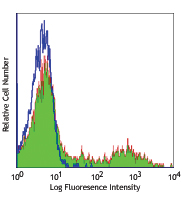
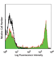
Compare Data Across All Formats
This data display is provided for general comparisons between formats.
Your actual data may vary due to variations in samples, target cells, instruments and their settings, staining conditions, and other factors.
If you need assistance with selecting the best format contact our expert technical support team.


Follow Us