 Login / Register


- Clone
- HCD56 (See other available formats)
- Regulatory Status
- RUO
- Other Names
- Leu-19, NKH1
- Isotype
- Mouse IgG1, κ
- Ave. Rating
- Submit a Review
- Product Citations
- publications

-

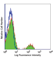
Human peripheral blood lymphocytes stained with HCD56 FITC
| Cat # | Size | Price | Quantity Check Availability | Save | ||
|---|---|---|---|---|---|---|
| 318303 | 25 tests | 85€ | ||||
| 318304 | 100 tests | 169€ | ||||
CD56 is a single transmembrane glycoprotein also known as NCAM (Neural Cell Adhesion Molecule), Leu-19, or NKH1. It is a member of the Ig superfamily. The 140 kD isoform is expressed on NK cells and NK-T cells. CD56 is also expressed in the brain (cerebellum and cortex) and at neuromuscular junctions. Certain large granular lymphocyte (LGL) leukemias, small-cell lung carcinomas, neuronal derived tumors, myelomas, and myeloid leukemias also express CD56. CD56 plays a role in homophilic and heterophilic adhesion via binding to itself or heparin sulfate.
Product DetailsProduct Details
- Reactivity
- Human
- Antibody Type
- Monoclonal
- Host Species
- Mouse
- Formulation
- Phosphate-buffered solution, pH 7.2, containing 0.09% sodium azide and BSA (origin USA)
- Preparation
- The antibody was purified by affinity chromatography, and conjugated with FITC under optimal conditions.
- Concentration
- Lot-specific (to obtain lot-specific concentration and expiration, please enter the lot number in our Certificate of Analysis online tool.)
- Storage & Handling
- The antibody solution should be stored undiluted between 2°C and 8°C, and protected from prolonged exposure to light. Do not freeze.
- Application
-
FC - Quality tested
- Recommended Usage
-
Each lot of this antibody is quality control tested by immunofluorescent staining with flow cytometric analysis. For flow cytometric staining, the suggested use of this reagent is 5 µl per million cells in 100 µl staining volume or 5 µl per 100 µl of whole blood.
- Excitation Laser
-
Blue Laser (488 nm)
- Application Notes
-
Clone HCD56 is not recommended for immunohistochemistry formalin-fixed paraffin-embedded tissue.
- Application References
-
- Kishimoto T, et al. Eds. 1997. Leucocyte Typing VI. Garland Publishing Inc. London.
- Correia DV, et al. 2011. Blood 118:992. (FC) PubMed
- Product Citations
- RRID
-
AB_604091 (BioLegend Cat. No. 318303)
AB_604100 (BioLegend Cat. No. 318304)
Antigen Details
- Structure
- Ig superfamily, single transmembrane or GPI-anchored glycoprotein
- Distribution
-
NK cells, T subset, neural tissue, some LGL and myeloid leukemias
- Function
- Adhesion
- Ligand/Receptor
- Heparin sulfate
- Cell Type
- B cells, Leukemia, Mesenchymal Stem Cells, Neurons, NK cells, T cells
- Biology Area
- Cell Adhesion, Cell Biology, Costimulatory Molecules, Immunology, Innate Immunity, Neuroscience, Stem Cells, Synaptic Biology
- Molecular Family
- Adhesion Molecules, CD Molecules
- Antigen References
-
1. Lanier L, et al. 1991. J. Immunol. 146:4421.
2. Hemperly J, et al. 1990. J. Mol. Neurosci. 2:71.
3. Cremer H, et al. 1994. Nature 367:455. - Gene ID
- 4684 View all products for this Gene ID
- UniProt
- View information about CD56 on UniProt.org
Related Pages & Pathways
Pathways


Customers Also Purchased
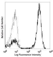
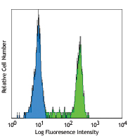
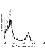
Compare Data Across All Formats
This data display is provided for general comparisons between formats.
Your actual data may vary due to variations in samples, target cells, instruments and their settings, staining conditions, and other factors.
If you need assistance with selecting the best format contact our expert technical support team.


Follow Us