 Login/Register


- Clone
- W3/25 (See other available formats)
- Regulatory Status
- RUO
- Other Names
- T4, L3T4, W3/25
- Isotype
- Mouse IgG1, κ
- Ave. Rating
- Submit a Review
- Product Citations
- publications

-

LOU rat splenocytes stained with W3/25 Alexa Fluor® 488 -

Rat frozen spleen section was fixed with 4% paraformaldehyde (PFA) for ten minutes at room temperature and blocked with 5% FBS for 30 minutes at room temperature. Then the section was stained with 10 µg/mL of CD4 (clone W3/25) Alexa Fluor® 488 (green), 10 µg/mL of CD8a (clone OX-8) Alexa Fluor® 647 (blue), and 10 µg/mL of purified CD38 (clone 14.27) overnight at 4°C, followed by 5 µg/mL of anti-mouse IgG2b (clone RMG2b-1) Alexa Fluor® 594 (red) for two hours at room temperature. The image was captured by 10X objective.
| Cat # | Size | Price | Quantity Check Availability | Save | ||
|---|---|---|---|---|---|---|
| 201511 | 25 µg | $101 | ||||
CD4 is a 55 kD glycoprotein also known as T4. Rat CD4 is a member of the immunoglobulin superfamily and is expressed on majority of thymocytes, macrophages, and a peripheral T cell subset (T helper cells). CD4 is a T cell co-receptor that interacts with the MHC class II molecule and is involved in T cell activation.
Product DetailsProduct Details
- Verified Reactivity
- Rat
- Antibody Type
- Monoclonal
- Host Species
- Mouse
- Immunogen
- Rat thymocyte membrane glycoproteins
- Formulation
- Phosphate-buffered solution, pH 7.2, containing 0.09% sodium azide.
- Preparation
- The antibody was purified by affinity chromatography and conjugated with Alexa Fluor® 488 under optimal conditions.
- Concentration
- 0.5 mg/ml
- Storage & Handling
- The antibody solution should be stored undiluted between 2°C and 8°C, and protected from prolonged exposure to light. Do not freeze.
- Application
-
FC - Quality tested
IHC-F - Verified - Recommended Usage
-
Each lot of this antibody is quality control tested by immunofluorescent staining with flow cytometric analysis. For flow cytometric staining, the suggested use of this reagent is ≤0.25 µg per million cells in 100 µl volume. For immunohistochemistry staining on frozen tissue sections, a concentration range of 2.5 - 10 µg/ml is suggested. It is recommended that the reagent be titrated for optimal performance for each application.
* Alexa Fluor® 488 has a maximum emission of 519 nm when it is excited at 488 nm.
Alexa Fluor® and Pacific Blue™ are trademarks of Life Technologies Corporation.
View full statement regarding label licenses - Excitation Laser
-
Blue Laser (488 nm)
- Application Notes
-
The W3/25 antibody has been shown to inhibit IL-2 production by T helper cells and to prevent autoimmune T cell transfer in an MBP induced EAE model in vivo. Additional reported applications (for the relevant formats) include: immunohistochemistry of acetone-fixed frozen sections1,2, inhibition of IL-2 production3, inhibition of MBP-induced T cell activation in EAE transfer model3.
-
Application References
(PubMed link indicates BioLegend citation) -
- Whiteland JL, et al. 1995. J. Histochem. Cytochem. 43:313. (IHC)
- Shioji K, et al. 2001. Circulation Res. 89:540. (IHC)
- Mannie MD, et al. 1993. J. Immunol. 151:7293.
- Kurtz CC, et al. 2007. Dev. Comp. Immunol. 31:415. PubMed
- Product Citations
-
- RRID
-
AB_572038 (BioLegend Cat. No. 201511)
Antigen Details
- Structure
- Ig superfamily, 55 kD
- Distribution
-
Majority of thymocytes, macrophages, and a peripheral T cell subset (T helper cells)
- Function
- T cell co-receptor, T cell activation
- Ligand/Receptor
- MHC class II molecule
- Cell Type
- Macrophages, T cells, Thymocytes
- Biology Area
- Immunology
- Molecular Family
- CD Molecules
- Antigen References
-
1. Brideau RJ, et al. 1980. Eur. J. Immunol. 10:609.
2. Clark SJ, et al. 187. P. Natl. Acad. Sci. USA 84:1649. - Gene ID
- 24933 View all products for this Gene ID
- UniProt
- View information about CD4 on UniProt.org
Related Pages & Pathways
Pathways


Other Formats
View All CD4 Reagents Request Custom Conjugation| Description | Clone | Applications |
|---|---|---|
| Purified anti-rat CD4 | W3/25 | FC,IHC-F |
| FITC anti-rat CD4 | W3/25 | FC |
| PE anti-rat CD4 | W3/25 | FC |
| APC anti-rat CD4 | W3/25 | FC |
| Alexa Fluor® 488 anti-rat CD4 | W3/25 | FC,IHC-F |
| APC/Cyanine7 anti-rat CD4 | W3/25 | FC |
| PE/Cyanine7 anti-rat CD4 | W3/25 | FC |
| PerCP/Cyanine5.5 anti-rat CD4 | W3/25 | FC |
Customers Also Purchased
Compare Data Across All Formats
This data display is provided for general comparisons between formats.
Your actual data may vary due to variations in samples, target cells, instruments and their settings, staining conditions, and other factors.
If you need assistance with selecting the best format contact our expert technical support team.
-
Purified anti-rat CD4

Lou rat splenocytes stained with purified W3/25, followed by... -
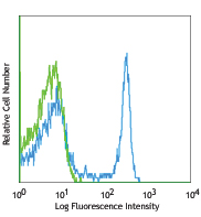
FITC anti-rat CD4

LOU rat splenocytes stained with W3/25 FITC -
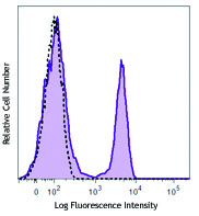
PE anti-rat CD4

LOU rat splenocytes stained with W3/25 PE -
APC anti-rat CD4

LOU rat splenocytes stained with W3/25 APC -
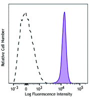
Alexa Fluor® 488 anti-rat CD4

LOU rat splenocytes stained with W3/25 Alexa Fluor® 488 
Rat frozen spleen section was fixed with 4% paraformaldehyde... -
APC/Cyanine7 anti-rat CD4

LOU rat splenocytes stained with W3/25 APC/Cyanine7 -
PE/Cyanine7 anti-rat CD4

LOU rat splenocytes stained with W3/25 PE/Cyanine7 -
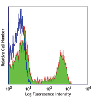
PerCP/Cyanine5.5 anti-rat CD4

LOU rat splenocytes were stained with CD3 (clone 1F4) PE and...


Follow Us