 Login/Register


- Clone
- APB5 (See other available formats)
- Regulatory Status
- RUO
- Other Names
- PDGF receptor-β, PDGFR-β, platelet-derived growth factor receptor beta
- Isotype
- Rat IgG2a, κ
- Ave. Rating
- Submit a Review
- Product Citations
- publications

-

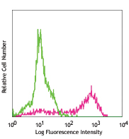
Mouse fibroblast NIH/3T3 cells stained with biotinylated APB5, followed by Sav-PE
| Cat # | Size | Price | Quantity Check Availability | Save | ||
|---|---|---|---|---|---|---|
| 136009 | 25 µg | $59 | ||||
| 136010 | 100 µg | $165 | ||||
Platelet-derived growth factor receptor-β (PDGFR-β), CD140b, is one of two receptors for platelet-derived growth factors (PDGFs) and binds to all isoforms of PDGFs. PDGFRβ is a receptor tyrosine kinase that forms homodimers or heterodimers on the surface upon ligand binding and phosphorylates substrates. PDGFRs consist of either homodimers of α/α, β/β, or heterodimers of α/β. PDGF receptors, α and β, are single glycoproteins with intracellular tyrosine kinase domain. Their ligand, PDGF, is a mitogen for connective tissue and glial cells. CD140b is expressed on embryonic tissues and mesenchymal-derived cells adult mice. PDGF plays a role in wound healing and acts as a chemoattractant for fibroblasts, smooth muscle cells, glial cells, monocytes, and neutrophils.
Product DetailsProduct Details
- Verified Reactivity
- Mouse
- Antibody Type
- Monoclonal
- Host Species
- Rat
- Immunogen
- Mouse PDGFR-β-hIgG1 recombinant fusion protein
- Formulation
- Phosphate-buffered solution, pH 7.2, containing 0.09% sodium azide.
- Preparation
- The antibody was purified by affinity chromatography, and conjugated with biotin under optimal conditions.
- Concentration
- 0.5 mg/ml
- Storage & Handling
- The antibody solution should be stored undiluted between 2°C and 8°C. Do not freeze.
- Application
-
FC - Quality tested
- Recommended Usage
-
Each lot of this antibody is quality control tested by immunofluorescent staining with flow cytometric analysis. For flow cytometric staining, the suggested use of this reagent is ≤ 1.0 µg per 106 cells in 100 µl volume. It is recommended that the reagent be titrated for optimal performance for each application.
- Application Notes
-
Additional reported (for the relevant formats) applications include: Western blotting and blocking function2. The LEAF™ purified antibody is recommended for functional assays.
-
Application References
(PubMed link indicates BioLegend citation) -
- Sano H, et al. 2001. Circulation 103:2955.
- Sano H, et al. 2002. Am. J. Pathol. 161:135. (Block)
- Product Citations
-
- RRID
-
AB_2236916 (BioLegend Cat. No. 136009)
AB_2236916 (BioLegend Cat. No. 136010)
Antigen Details
- Structure
- Beta chain of the platelet-derived growth factor receptor, a receptor tyrosine kinase that forms homo- or hetero-dimers on the surface after ligand binding.
- Distribution
-
Expressed on embryonic tissues and mesenchymal-derived cells of adult mouse.
- Function
- Play a role in wound healing and act as a chemoattractant for fibroblasts, smooth muscle cells, glial cells, monocytes and neutrophils.
- Ligand/Receptor
- PDGFs
- Cell Type
- Embryonic Stem Cells, Mesenchymal Stem Cells
- Biology Area
- Cell Biology, Immunology, Neuroscience, Neuroscience Cell Markers, Stem Cells
- Molecular Family
- CD Molecules, Cytokine/Chemokine Receptors
- Antigen References
-
1. Soriano P, et al. 1994. Genes Dev. 8:1888
2. Takakura N, et al. 1996. J Invest Dermatol. 107:770
3. Yarden Y, et al. 1986. Nature 323:226 - Gene ID
- 18596 View all products for this Gene ID
- UniProt
- View information about CD140b on UniProt.org
Related Pages & Pathways
Pathways


Related FAQs
- How many biotin molecules are per antibody structure?
- We don't routinely measure the number of biotins with our antibody products but the number of biotin molecules range from 3-6 molecules per antibody.
Other Formats
View All CD140b Reagents Request Custom Conjugation| Description | Clone | Applications |
|---|---|---|
| Purified anti-mouse CD140b | APB5 | FC,WB,Block |
| PE anti-mouse CD140b | APB5 | FC |
| APC anti-mouse CD140b | APB5 | FC |
| Biotin anti-mouse CD140b | APB5 | FC |
| Ultra-LEAF™ Purified anti-mouse CD140b | APB5 | FC,Block,WB |
Customers Also Purchased
Compare Data Across All Formats
This data display is provided for general comparisons between formats.
Your actual data may vary due to variations in samples, target cells, instruments and their settings, staining conditions, and other factors.
If you need assistance with selecting the best format contact our expert technical support team.
-
Purified anti-mouse CD140b

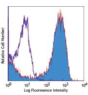
Mouse fibroblast NIH/3T3 cells stained with APB5 PE 
C57BL/6 splencoytes stained with APB5 PE -
PE anti-mouse CD140b
Mouse fibroblast NIH/3T3 cells stained with APB5 PE
C57BL/6 splencoytes stained with APB5 PE -
APC anti-mouse CD140b

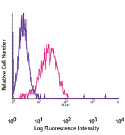
Mouse fibroblast NIH/3T3 cells stained with APB5 APC -
Biotin anti-mouse CD140b

Mouse fibroblast NIH/3T3 cells stained with biotinylated APB... -
Ultra-LEAF™ Purified anti-mouse CD140b


Follow Us