 Login/Register


- Clone
- Poly5272 (See other available formats)
- Regulatory Status
- RUO
- Other Names
- TNF ligand-related molecule 1 (TL1), TL1A, vascular endothelial cell growth inhibitor (V)
- Isotype
- Goat Polyclonal IgG
- Ave. Rating
- Submit a Review
- Product Citations
- publications

-
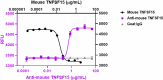
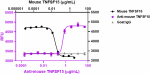
Recombinant mouse TNFSF15 (Cat. No. 753006) (black circles) induces apoptotic death on human TF-1 cells. LEAF™ purified anti-mouse TNFSF15 (clone Poly5272) (purple squares) neutralizes the cell apoptotic death induced by recombinant mouse TNFSF15 at 0.2 µg/mL on human TF-1 cells in a dose-dependent manner, whereas the LEAF™ purified goat IgG isotype control (gray triangles) does not have this effect. ND50 range: 0.2 - 1.8 µg/mL. -

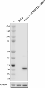
Total cell lysates (15 µg total protein) from indicated cell lines spiked with or without 20 ng of recombinant mouse TNFSF15 protein (Cat. No. 753002) were resolved by 4-12 % Bis-Tris gel electrophoresis, transferred to a PVDF membrane, and probed with 0.1 µg/mL of LEAF™ purified anti-mouse TNFSF15 (clone Poly5272) overnight at 4°C. Proteins were visualized by chemiluminescence detection using HRP-conjugated anti-goat IgG. Direct-Blot™ HRP anti-GAPDH (Cat. No. 607904) was used as a loading control at a 1:50000 dilution. Lane M: Molecular weight ladder -
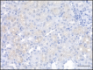
IHC staining on formalin-fixed paraffin-embedded human kidney tissue. Following antigen retrieval using 1X Sodium Citrate H.I.E.R. (Cat. No. 928502), the tissue was incubated with 5 µg/mL of LEAF™ purified anti-mouse TNFSF15 (clone Poly5272) overnight at 4°C. Ultra-Streptavidin HRP Kit (Multi-Species, DAB, Cat. No. 929501) and HRP-conjugated anti-goat IgG were used for detection followed by hematoxylin counterstaining, according to the protocol provided. The image was captured with a 40X objective. Scale bar: 50 µm
| Cat # | Size | Price | Quantity Check Availability | Save | ||
|---|---|---|---|---|---|---|
| 527203 | 100 µg | $375 | ||||
TNFSF15 is member 15 of the tumor necrosis factor (ligand) superfamily and also is a type II transmembrane protein. It is expressed as a membrane-bound protein and later released as a soluble protein via the ectodomain shedding by TNF-α converting enzyme (TACE). TNFSF15 exhibits approximately 20-30% homology to other TNFSF members. The functional receptor of TNFSF15 is the death receptor 3 (DR3). The engagement of this receptor on T cells by TNFSF15 expressed on dendritic cells (DCs) triggers a costimulatory signal in the T cells that induces IFN-γ production via NF-κB. DR3+ tumor cell lines treated with soluble TNFSF15 in the presence of cycloheximide results in caspase-dependent apoptosis. The TNFSF15-DR3 interaction is inhibited by the DcR3 decoy receptor. DcR3 also binds to LIGHT (TNFSF14) and FasL (TNFSF6). TNFSF15, in conjunction with IL-12, IL-15, and IL-18, induces the expression of co-stimulatory molecules CD40L (CD154) and OX40 (CD134) on activated CD4 T cells. The co-stimulatory process is associated with the expression of IL-2Rα (CD25) and α-chain of LFA-1 (CD11a) on CD4 T cells. Also, the costimulation with TNFSF15 induces IL-22 and GM-CSF. In addition, TNFSF15, in concert with IL-12, IL-15, and IL-18, induces the production of IL-6 and TNF-α from leukocytes of healthy donors. TNFSF15 has been associated with inflammatory bowel disease (IBD), and its expression correlates with the severity of intestinal inflammation and increased IFN-γ production in intestinal lamina propria. In addition, TNFSF15 (produced by lamina propria macrophages) induces Th1 and Th17 immune responses in cooperation with IL-23 in patients with Crohn's disease. TNFSF15 is also related to rheumatoid arthritis (RA), ankylosing spondylitis, and psoriasis.
Product DetailsProduct Details
- Verified Reactivity
- Mouse
- Antibody Type
- Polyclonal
- Host Species
- Goat
- Immunogen
- Recombinant mouse TNFSF15
- Formulation
- 0.2 µm filtered in phosphate-buffered solution, pH 7.2, containing no preservative.
- Endotoxin Level
- Less than 0.1 EU/µg of the protein (< 0.01 ng/µg of the protein) as determined by the LAL test.
- Preparation
- The LEAF™ (Low Endotoxin, Azide-Free) antibody was purified by affinity chromatography.
- Concentration
- Lot-specific (to obtain lot-specific concentration and expiration, please enter the lot number in our Certificate of Analysis online tool.)
- Storage & Handling
- Upon receipt, store frozen at -20°C. Make small volume aliquots if needed and avoid repeated freeze-thaw cycles to prevent denaturing the antibody.
- Application
-
Neut - Quality tested
WB, IHC-P - Verified - Recommended Usage
-
Each lot of this antibody is quality control tested by neutralizing apoptosis induced by recombinant mouse TNFSF15 (Cat. No. 753006) at 0.2 µg/mL on human TF-1 cells. ND50 range: 0.2 - 1.8 µg/mL. For western blotting, the suggested use of this reagent is 0.02 - 0.5 µg/mL. For immunohistochemistry on formalin-fixed paraffin-embedded tissue sections, a concentration range of 5.0 - 10 µg/mL is suggested. It is recommended that the reagent be titrated for optimal performance for each application.
- RRID
-
AB_2941597 (BioLegend Cat. No. 527203)
Antigen Details
- Structure
- Homotrimer
- Distribution
-
Endothelial cells, endothelial progenitor cells, monocytes, monocyte-derived dendritic cells (DCs), synovial fibroblast-like cells, and CD4 and CD8 lymphocytes
- Function
- TNFSF15 acts as a costimulatory signal inducing T cell proliferation and secretion of IFN-γ and GM-CSF. Its expression is induced by TNF-α, IL-1-α, and PMA.
- Interaction
- Activated T cells, natural killer (NK) cells, NKT cells, macrophages, and endothelial cells
- Ligand/Receptor
- DR3 (TNFRSF25) and decoy receptor TR6/DcR3 (TNFRSF6B)
- Cell Type
- Dendritic cells, Endothelial cells, Monocytes, T cells
- Biology Area
- Immunology, Innate Immunity
- Molecular Family
- Cytokines/Chemokines
- Antigen References
-
- Migone TS, et al. 2002. Immunity. 16:479-92.
- Bamias G, et al. 2003. J Immunol. 171:4868-74.
- Kamada N, et al. 2010. Inflamm Bowel Dis. 16:568-75.
- Aiba Y & Nakamura M. 2013. Mediators Inflamm. 2013:258164.
- Reichwald K, et al. 2014. PLoS One. 9(8):e105627.
- Reichwald K, et al. 2014. PLoS One. 9(1):e85793.
- Gene ID
- 326623 View all products for this Gene ID
- UniProt
- View information about TNFSF15 on UniProt.org
Related Pages & Pathways
Pathways


Related FAQs
- Do you guarantee that your antibodies are totally pathogen free?
-
BioLegend does not test for pathogens in-house aside from the GoInVivo™ product line. However, upon request, this can be tested on a custom basis with an outside, independent laboratory.
- Does BioLegend test each Ultra-LEAF™ antibody by functional assay?
-
No, BioLegend does not test Ultra-LEAF™ antibodies by functional assays unless otherwise indicated. Due to the possible complexities and variations of uses of biofunctional antibodies in different assays and because of the large product portfolio, BioLegend does not currently perform functional assays as a routine QC for the antibodies. However, we do provide references in which the antibodies were used for functional assays and we do perform QC to verify the specificity and quality of the antibody based on our strict specification criteria.
- Does BioLegend test each Ultra-LEAF™ antibody for potential pathogens?
-
No, BioLegend does not test for pathogens in-house unless otherwise indicated. However, we can recommend an outside vendor to perform this testing as needed.
- Have you tested this Ultra-LEAF™ antibody for in vivo or in vitro applications?
-
We don't test our antibodies for in vivo or in vitro applications unless otherwise indicated. Depending on the product, the TDS may describe literature supporting usage of a particular product for bioassay. It may be best to further consult the literature to find clone specific information.
Other Formats
View All TNFSF15 Reagents Request Custom Conjugation| Description | Clone | Applications |
|---|---|---|
| LEAF™ Purified anti-mouse TNFSF15 | Poly5272 | Neut,WB,IHC-P |
Compare Data Across All Formats
This data display is provided for general comparisons between formats.
Your actual data may vary due to variations in samples, target cells, instruments and their settings, staining conditions, and other factors.
If you need assistance with selecting the best format contact our expert technical support team.


Follow Us