 Login/Register


- Clone
- P-syn/81A (See other available formats)
- Regulatory Status
- RUO
- Other Names
- Synuclein alpha-140, non-A4 component of amyloid, alpha-synuclein, isoform NACP140, non-A beta component of AD amyloid Parkinson disease (autosomal dominant, Lewy body) 4
- Previously
-
Covance Catalog# MMS-5091
- Isotype
- Mouse IgG2a, κ
- Ave. Rating
- Submit a Review
- Product Citations
- publications

-

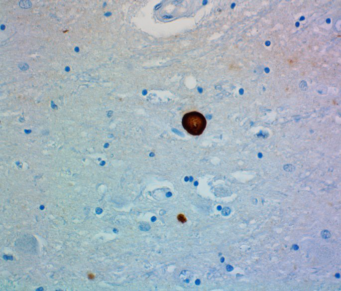
IHC staining of purified anti-α-Synuclein Phospho (Ser129) antibody (clone P-syn/81A) on formalin-fixed paraffin-embedded diseased human brain tissue. Following antigen retrieval using 70% formic acid for 20 minutes at room temperature, the tissue was incubated with 1 µg/ml of the primary antibody overnight at 4°C. BioLegend´s Ultra-Streptavidin (USA) HRP kit (Multi-Species, DAB, Cat. No. 929901) was used for detection followed by hematoxylin counterstaining, according to the protocol provided. The image was captured with a 40X objective.
| Cat # | Size | Price | Quantity Check Availability | Save | ||
|---|---|---|---|---|---|---|
| 825702 | 25 µL | $129 | ||||
| 825701 | 100 µL | $335 | ||||
α-synuclein is expressed principally in the nervous system, but it is also produced in other tissues, including the skin. In the brain, the protein is primarily neuronal, but it is also present in glia. Neuronal α-synuclein is concentrated in presynapt ic nerve terminals, interacts with plasma membrane phospholipids, and is also present in nuclei and mitochondria. At least three isoforms of α-synuclein are produced through alternative splicing. The most common isoform is a 140 amino acid-long transcript. Other isoforms includes, a-synuclein-126, lacking residues 41-54; and α-synuclein-112, which lacks residues 103-130. α-synuclein’s physiological role is poorly understood, but the protein has been implicated in regulating dopamine release and transport, synaptic vesicle clustering, and functioning as a SNARE-complex chaperone. α-synuclein fibrils are a major component of the intracellular Lewy bodies that are associated with Parkinson's disease, Lewy body dementia, and multiple system atrophy. α-synuclein is phosphorylated at low levels under normal physiological conditions whereas the majority of the protein is phosphorylated in Lewy bodies at S129.
Product DetailsProduct Details
- Verified Reactivity
- Human
- Antibody Type
- Monoclonal
- Host Species
- Mouse
- Immunogen
- This monoclonal antibody was raised against a synthetic peptide corresponding to amino acids 124 - 134 of α-synuclein, phosphorylated at Serine 129, and conjugated to KLH via a C-terminal Cysteine.
- Formulation
-
Phosphate-buffered solution with 0.09% azide.
Previous lots of this product may have been formulated with 0.1% or 0.05% NaN3 instead of 0.09% NaN3. For further information please contact BioLegend Technical Support or Customer Service. - Preparation
- The antibody was purified by affinity chromatography.
- Concentration
- 1.0 mg/ml
- Storage & Handling
- The antibody solution should be stored undiluted between 2°C and 8°C. Do not freeze.
- Application
-
IHC-P - Quality tested
ICC, IHC-F, WB - Reported in the literature, not verified in house
SB - Community verified - Recommended Usage
-
Each lot of this antibody is quality control tested by formalin-fixed paraffin-embedded immunohistochemical staining. For immunohistochemistry, a concentration range of 1 - 2 µg/ml is suggested. It is recommended that the reagent be titrated for optimal performance for each application.
- Application Notes
-
This antibody is effective in immunohistochemistry (IHC-P). Additional reported applications (for the relevant formats) include: Western blotting, immunohistochemisty on frozen tissue sections (IHC-F), and immunocytochemistry.
P-syn/81A is a mAb that is specific to alpha synuclein that has been phosphorylated on serine 129. - Additional Product Notes
-
This product has been verified for IHC-P (Immunohistochemistry- formalin-fixed paraffin-embedded tissues) on the NanoString GeoMx® Digital Spatial Profiler. The GeoMx® enables researchers to perform spatial analysis of protein and RNA targets in FFPE and fresh frozen human and mouse samples. For more information about our spatial biology products and the GeoMx® platform, please visit our spatial biology page.
-
Application References
(PubMed link indicates BioLegend citation) - Product Citations
-
- RRID
-
AB_2734593 (BioLegend Cat. No. 825702)
AB_2564891 (BioLegend Cat. No. 825701)
Antigen Details
- Structure
- α-synuclein's canonical isoform consists of 140 amino acids, which consist of four 11-residue repeats containing the consensus sequence KTKEGV. α-synuclein has an apparent molecular mass of 14 kD.
- Distribution
-
Tissue distribution: primarily nervous system, but lower expression in other tissues such as skin.
Cellular distribution: cytoskeleton, cytosol, lysosome, mitochondria, nucleus, plasma membrane, and extracellular. - Function
- The function of α-synuclein in the healthy brain is currently unknown.
- Biology Area
- Cell Biology, Neurodegeneration, Neuroscience, Protein Misfolding and Aggregation
- Molecular Family
- α-Synuclein, Phospho-Proteins
- Antigen References
- Gene ID
- 6622 View all products for this Gene ID
- UniProt
- View information about alpha-Synuclein Phospho Ser129 on UniProt.org

Related FAQs
- If an antibody clone has been previously successfully used in IBEX in one fluorescent format, will other antibody formats work as well?
-
It’s likely that other fluorophore conjugates to the same antibody clone will also be compatible with IBEX using the same sample fixation procedure. Ultimately a directly conjugated antibody’s utility in fluorescent imaging and IBEX may be specific to the sample and microscope being used in the experiment. Some antibody clone conjugates may perform better than others due to performance differences in non-specific binding, fluorophore brightness, and other biochemical properties unique to that conjugate.
- Will antibodies my lab is already using for fluorescent or chromogenic IHC work in IBEX?
-
Fundamentally, IBEX as a technique that works much in the same way as single antibody panels or single marker IF/IHC. If you’re already successfully using an antibody clone on a sample of interest, it is likely that clone will have utility in IBEX. It is expected some optimization and testing of different antibody fluorophore conjugates will be required to find a suitable format; however, legacy microscopy techniques like chromogenic IHC on fixed or frozen tissue is an excellent place to start looking for useful antibodies.
- Are other fluorophores compatible with IBEX?
-
Over 18 fluorescent formats have been screened for use in IBEX, however, it is likely that other fluorophores are able to be rapidly bleached in IBEX. If a fluorophore format is already suitable for your imaging platform it can be tested for compatibility in IBEX.
- The same antibody works in one tissue type but not another. What is happening?
-
Differences in tissue properties may impact both the ability of an antibody to bind its target specifically and impact the ability of a specific fluorophore conjugate to overcome the background fluorescent signal in a given tissue. Secondary stains, as well as testing multiple fluorescent conjugates of the same clone, may help to troubleshoot challenging targets or tissues. Using a reference control tissue may also give confidence in the specificity of your staining.
- How can I be sure the staining I’m seeing in my tissue is real?
-
In general, best practices for validating an antibody in traditional chromogenic or fluorescent IHC are applicable to IBEX. Please reference the Nature Methods review on antibody based multiplexed imaging for resources on validating antibodies for IBEX.
Other Formats
View All α-Synuclein Phospho (Ser12) Reagents Request Custom Conjugation| Description | Clone | Applications |
|---|---|---|
| Purified anti-α-Synuclein Phospho (Ser129) | P-syn/81A | IHC-P,ICC,IHC-F,WB,SB |
| Biotin anti-α-Synuclein Phospho (Ser129) | P-syn/81A | IHC-P |
| Alexa Fluor® 594 anti-α-Synuclein Phospho (Ser129) | P-syn/81A | IHC-P |
Customers Also Purchased
Compare Data Across All Formats
This data display is provided for general comparisons between formats.
Your actual data may vary due to variations in samples, target cells, instruments and their settings, staining conditions, and other factors.
If you need assistance with selecting the best format contact our expert technical support team.
-
Purified anti-α-Synuclein Phospho (Ser129)

IHC staining of purified anti-α-Synuclein Phospho (Ser129) a... -
Biotin anti-α-Synuclein Phospho (Ser129)

IHC staining of α-synuclein deposits with biotin anti-α-Synu... -
Alexa Fluor® 594 anti-α-Synuclein Phospho (Ser129)

IHC staining of Alexa Fluor® 594 anti-α-Synuclein Phospho (...


Follow Us